Copyright
©The Author(s) 2022.
World J Clin Cases. Jan 21, 2022; 10(3): 985-991
Published online Jan 21, 2022. doi: 10.12998/wjcc.v10.i3.985
Published online Jan 21, 2022. doi: 10.12998/wjcc.v10.i3.985
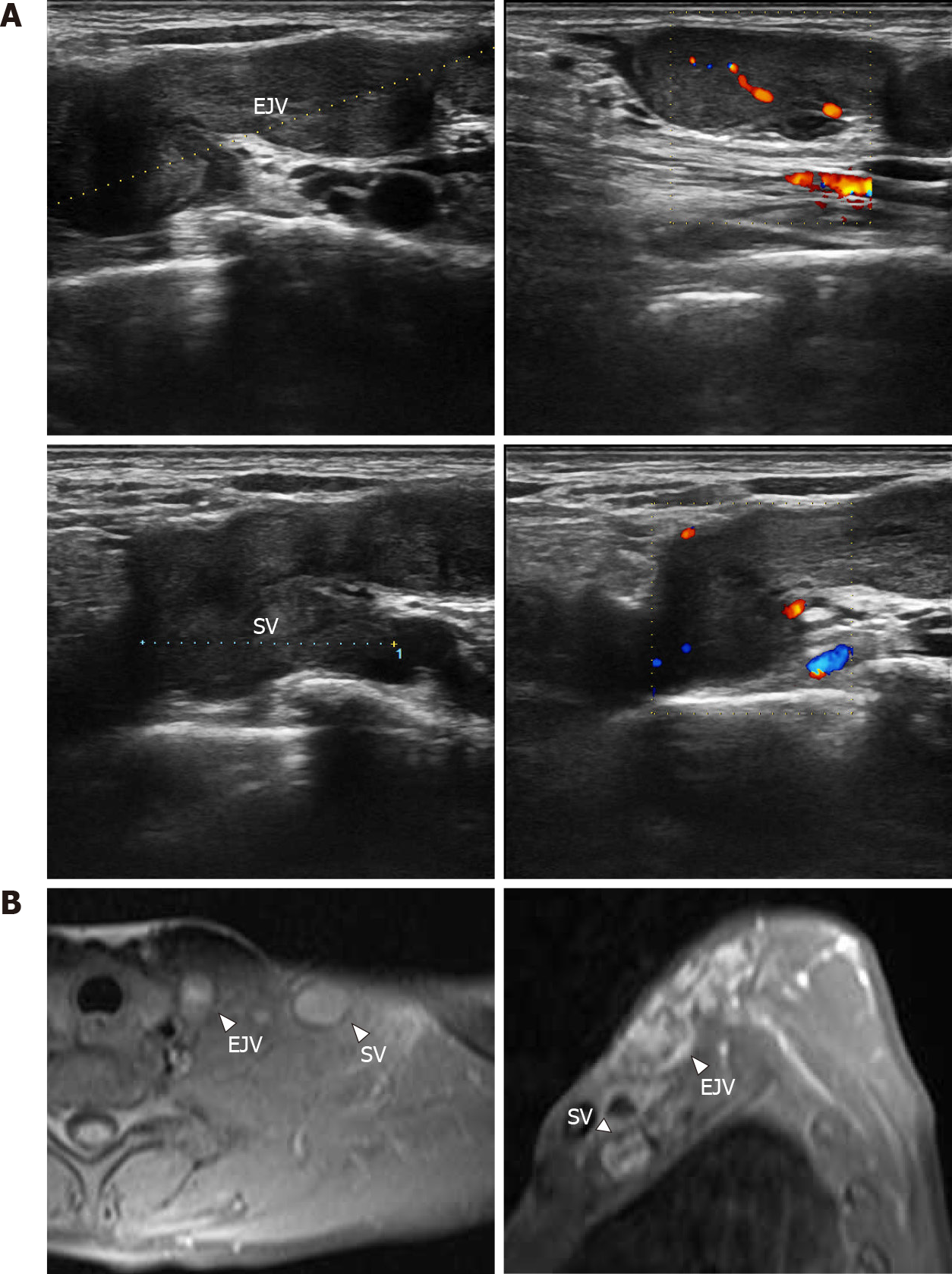
Figure 1 Imaging examination.
A: Duplex ultrasonography: a circular solid hypoechoic mass could be seen along the external jugular vein, with a general length of approximately 4.2 cm; there was no blood flow signal passing through the lumen, and the mass invaded into the subclavian vein along the external jugular vein. Hypoechoic masses were observed in some areas of the subclavian vein, involving a length of approximately 2.1 cm; B: Magnetic resonance imaging revealed an enhanced longitudinal mass-like lesion in the left supraclavicular fossa. EJV: External jugular vein; SV: Subclavian veins.
- Citation: Meng XH, Liu YC, Xie LS, Huang CP, Xie XP, Fang X. Intravascular fasciitis involving the external jugular vein and subclavian vein: A case report . World J Clin Cases 2022; 10(3): 985-991
- URL: https://www.wjgnet.com/2307-8960/full/v10/i3/985.htm
- DOI: https://dx.doi.org/10.12998/wjcc.v10.i3.985


