
















©The Author(s) 2022.
World J Clin Cases. Jan 21, 2022; 10(3): 811-819
Published online Jan 21, 2022. doi: 10.12998/wjcc.v10.i3.811
Published online Jan 21, 2022. doi: 10.12998/wjcc.v10.i3.811
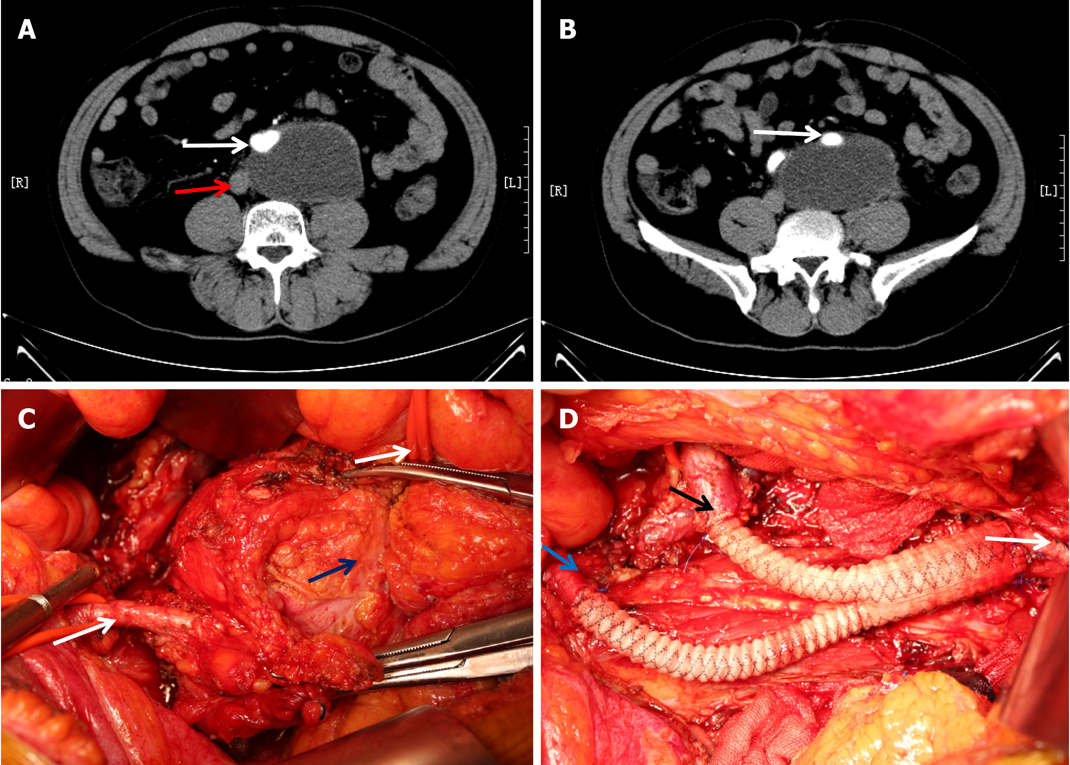
Figure 2 Computed tomography images of a case of myxofibrosarcoma and key steps in the surgery.
A: A layer of the arterial phase of the computed tomography scan indicated a retroperitoneal mass with a diameter of approximately 9 cm surrounding the abdominal aorta (white arrow) and adherent to the inferior vena cava (red arrow); B: Another layer indicated that the mass surrounded the left common iliac artery (white arrow); C: During surgery, the left external iliac artery (lower left arrow) and abdominal aorta (upper right arrow) were dissociated; the blue arrow indicates the mass; D: “Y-type” artificial vascular reconstruction of the abdominal aorta (white arrow), right common iliac artery (black arrow), and left external iliac artery (blue arrow) was performed after tumor removal.
- Citation: Li WX, Tong HX, Lv CT, Yang H, Zhao G, Lu WQ, Zhang Y. Management of retroperitoneal sarcoma involving the iliac artery: Single-center surgical experience. World J Clin Cases 2022; 10(3): 811-819
- URL: https://www.wjgnet.com/2307-8960/full/v10/i3/811.htm
- DOI: https://dx.doi.org/10.12998/wjcc.v10.i3.811