Copyright
©The Author(s) 2022.
World J Clin Cases. Jan 21, 2022; 10(3): 1106-1115
Published online Jan 21, 2022. doi: 10.12998/wjcc.v10.i3.1106
Published online Jan 21, 2022. doi: 10.12998/wjcc.v10.i3.1106
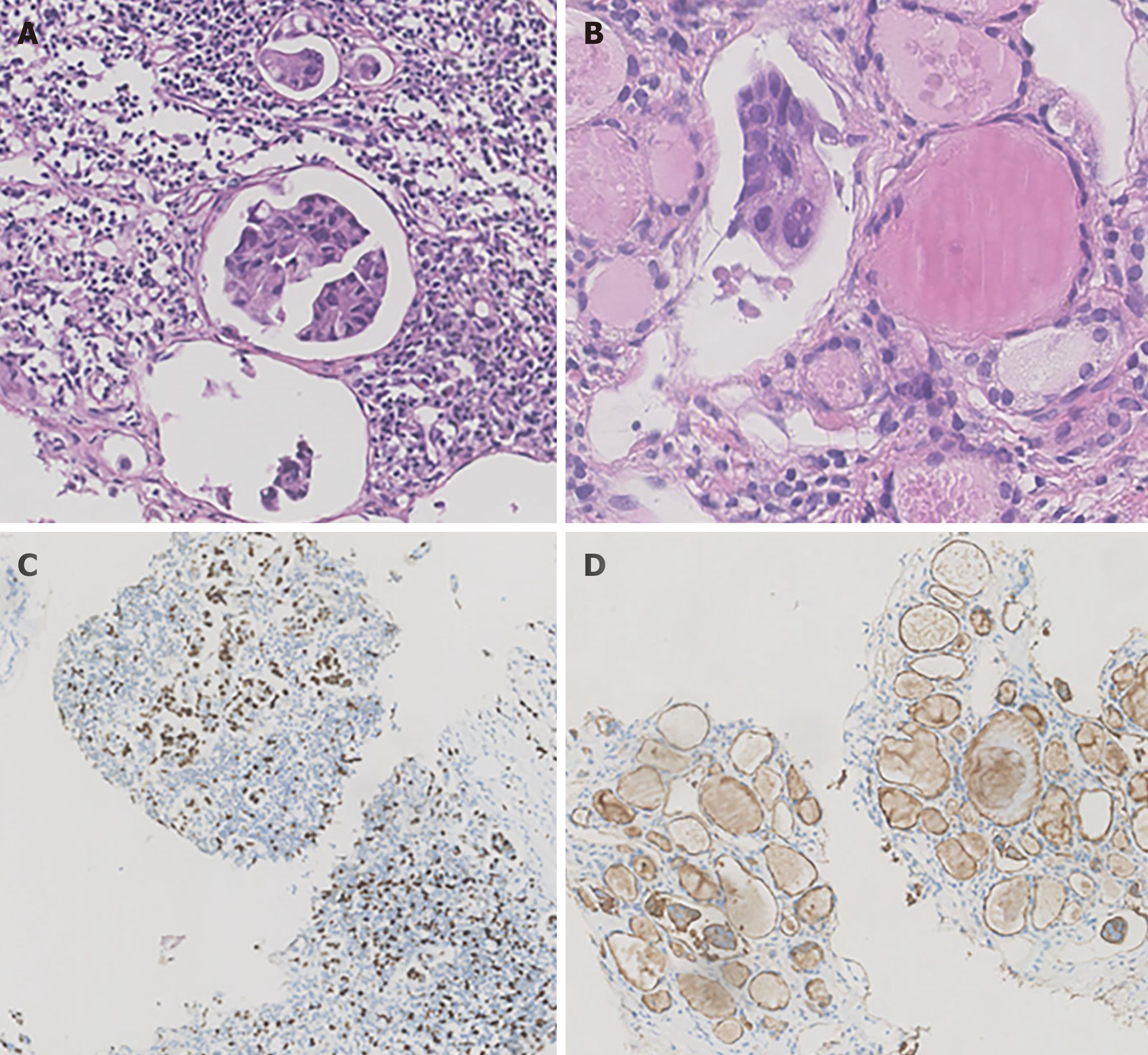
Figure 3 Hematoxylin-eosin staining and immunohistochemical staining of the left cervical lymph nodes and thyroid.
A: Atypical cells from a breast cancer metastatic to the left cervical lymph node processed with histology; B: Nested tumor cells mixed in the thyroid follicles from a breast cancer metastatic to the thyroid gland processed with histology; C: Immunocytochemical evaluation of the Ki-67 index in cervical lymph node metastatic breast carcinoma. The tumor cells are diffusely positive for Ki-67; D: Immunocytochemical evaluation of epithelial membrane antigen (EMA) in thyroid metastatic breast carcinoma. The tumor cells are diffusely positive for EMA.
- Citation: Wen W, Jiang H, Wen HY, Peng YL. Metastasis to the thyroid gland from primary breast cancer presenting as diffuse goiter: A case report and review of literature. World J Clin Cases 2022; 10(3): 1106-1115
- URL: https://www.wjgnet.com/2307-8960/full/v10/i3/1106.htm
- DOI: https://dx.doi.org/10.12998/wjcc.v10.i3.1106


