Copyright
©The Author(s) 2022.
World J Clin Cases. Oct 16, 2022; 10(29): 10763-10771
Published online Oct 16, 2022. doi: 10.12998/wjcc.v10.i29.10763
Published online Oct 16, 2022. doi: 10.12998/wjcc.v10.i29.10763
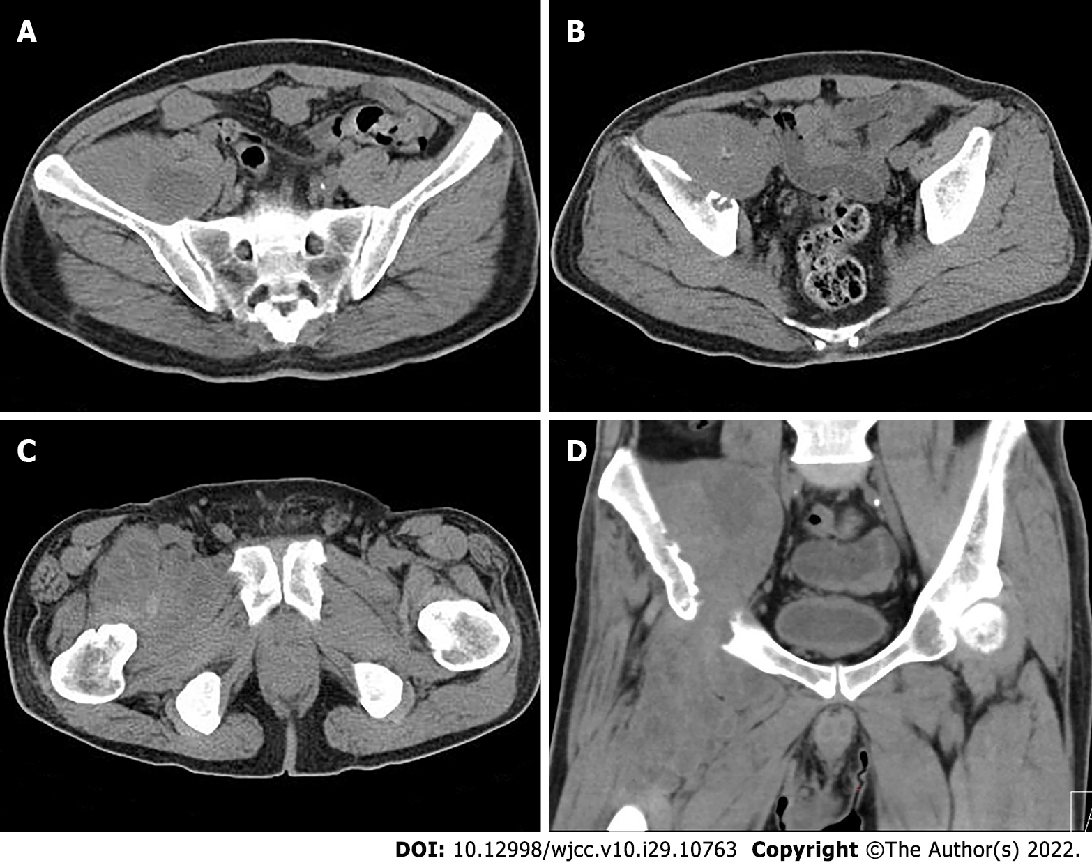
Figure 2 Computed tomography of the pelvis reveals a soft tissue mass in the intermuscular space anterior to the right iliopsoas muscle and the right upper femur.
A: The mass showed soft tissue density with lamellar hypodense necrosis inside; B: Osteolytic bone destruction of the right iliac bone was displayed clearly; C: The mass had poorly defined margins; and D: The massive tumor growed along the tendon.
- Citation: Huang WP, Gao G, Yang Q, Chen Z, Qiu YK, Gao JB, Kang L. Malignant giant cell tumors of the tendon sheath of the right hip: A case report. World J Clin Cases 2022; 10(29): 10763-10771
- URL: https://www.wjgnet.com/2307-8960/full/v10/i29/10763.htm
- DOI: https://dx.doi.org/10.12998/wjcc.v10.i29.10763


