Copyright
©The Author(s) 2022.
World J Clin Cases. Oct 16, 2022; 10(29): 10763-10771
Published online Oct 16, 2022. doi: 10.12998/wjcc.v10.i29.10763
Published online Oct 16, 2022. doi: 10.12998/wjcc.v10.i29.10763
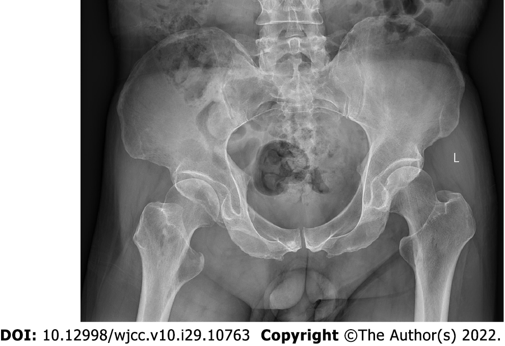
Figure 1 Pelvis radiography.
Multiple cystic hypodense lesions with variable sizes and well-defined borders were shown in the right iliac bone and right upper femur, suggesting osteolytic bone destruction.
- Citation: Huang WP, Gao G, Yang Q, Chen Z, Qiu YK, Gao JB, Kang L. Malignant giant cell tumors of the tendon sheath of the right hip: A case report. World J Clin Cases 2022; 10(29): 10763-10771
- URL: https://www.wjgnet.com/2307-8960/full/v10/i29/10763.htm
- DOI: https://dx.doi.org/10.12998/wjcc.v10.i29.10763


