
















©The Author(s) 2022.
World J Clin Cases. Oct 16, 2022; 10(29): 10565-10574
Published online Oct 16, 2022. doi: 10.12998/wjcc.v10.i29.10565
Published online Oct 16, 2022. doi: 10.12998/wjcc.v10.i29.10565
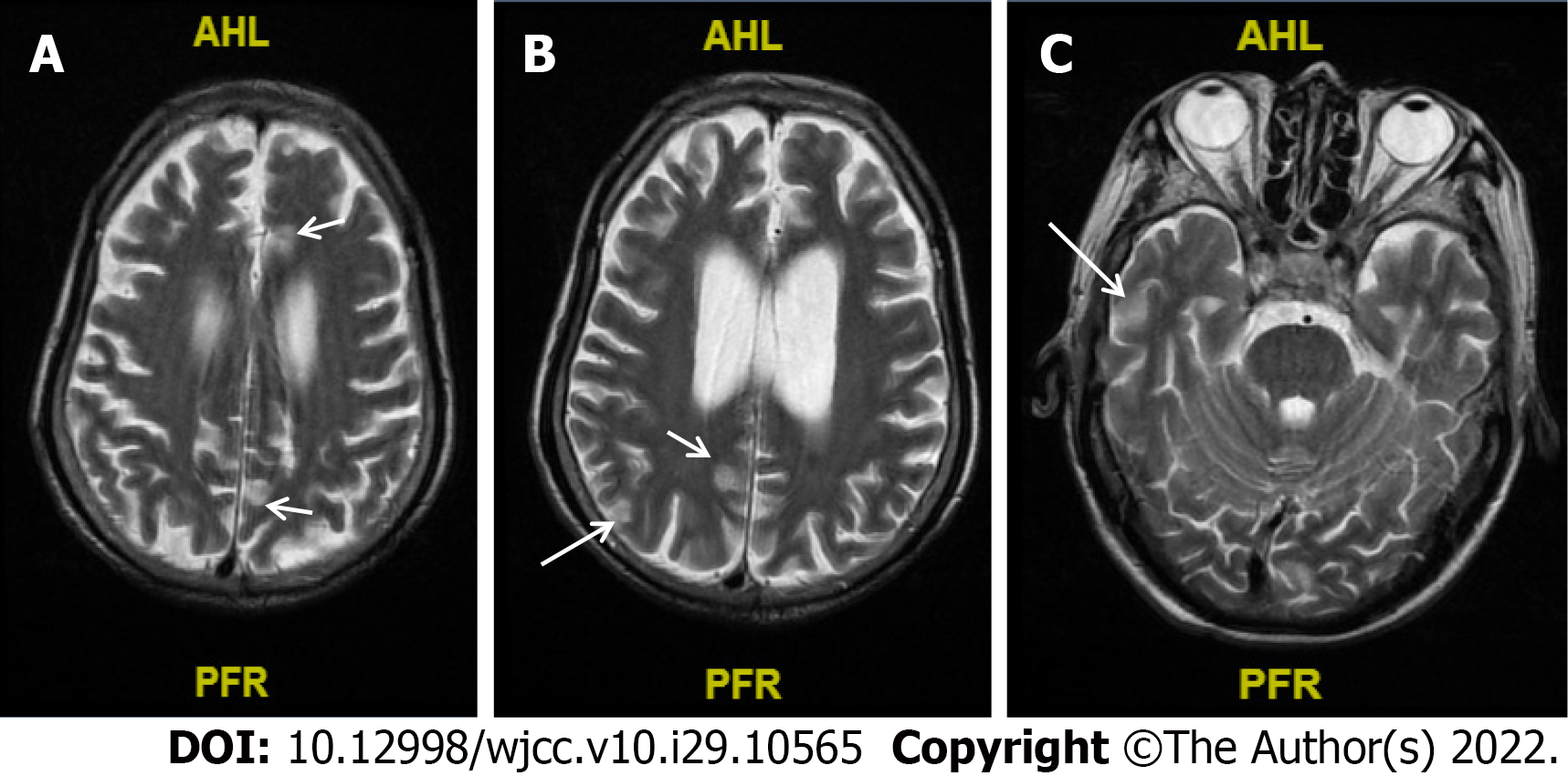
Figure 3 The result of cranial magnetic resonance upon the patient's second admission.
A-C: Abnormal signals diverged from the subfrontal cortex (A), midbrain (B), and the posterior horns of both sides of the ventricle (C) (arrow pointing), and no obvious brain abscess formation was noted.
- Citation: Wu GX, Zhou JY, Hong WJ, Huang J, Yan SQ. Treatment failure in a patient infected with Listeria sepsis combined with latent meningitis: A case report. World J Clin Cases 2022; 10(29): 10565-10574
- URL: https://www.wjgnet.com/2307-8960/full/v10/i29/10565.htm
- DOI: https://dx.doi.org/10.12998/wjcc.v10.i29.10565