Copyright
©The Author(s) 2022.
World J Clin Cases. Oct 6, 2022; 10(28): 10252-10259
Published online Oct 6, 2022. doi: 10.12998/wjcc.v10.i28.10252
Published online Oct 6, 2022. doi: 10.12998/wjcc.v10.i28.10252
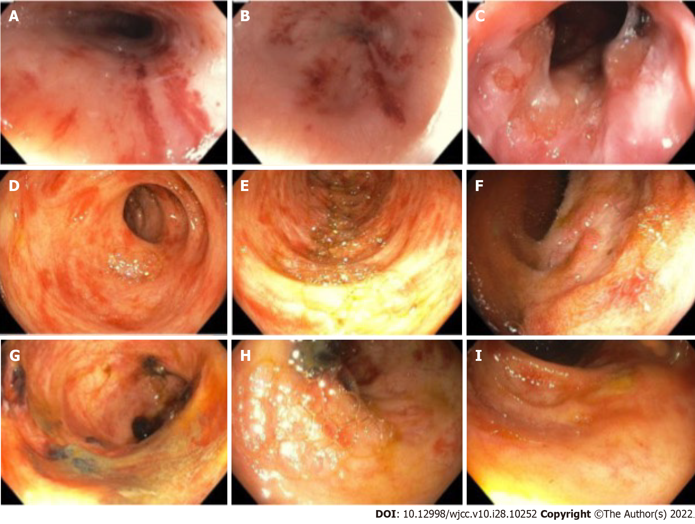
Figure 4 Esophagogastroduodenoscopy and colonoscopy images.
A-C: Esophagogastroduodenoscopy shows petechial lesions and friable mucosa scattered throughout distal esophagus to duodenum; D-I: Colonoscopy images show petechial lesions and shallow ulcers in colon.
- Citation: Bilton SE, Shah N, Dougherty D, Simpson S, Holliday A, Sahebjam F, Grider DJ. Persistent diarrhea with petechial rash - unusual pattern of light chain amyloidosis deposition on skin and gastrointestinal biopsies: A case report. World J Clin Cases 2022; 10(28): 10252-10259
- URL: https://www.wjgnet.com/2307-8960/full/v10/i28/10252.htm
- DOI: https://dx.doi.org/10.12998/wjcc.v10.i28.10252


