Copyright
©The Author(s) 2022.
World J Clin Cases. Sep 26, 2022; 10(27): 9945-9953
Published online Sep 26, 2022. doi: 10.12998/wjcc.v10.i27.9945
Published online Sep 26, 2022. doi: 10.12998/wjcc.v10.i27.9945
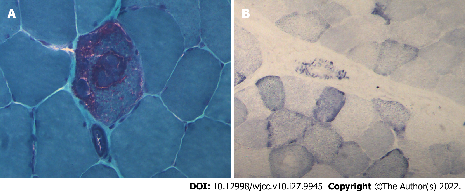
Figure 4 Muscle pathology images of case 1.
A: Modified gomori trichromatic staining showed red fiber breakage; B: Succinate dehydrogenase staining showed blue-stained fiber breakage and hyperstained small vessels (magnification: 200 ×).
- Citation: Yang X, Fu LJ. Familial mitochondrial encephalomyopathy, lactic acidosis, and stroke-like episode syndrome: Three case reports. World J Clin Cases 2022; 10(27): 9945-9953
- URL: https://www.wjgnet.com/2307-8960/full/v10/i27/9945.htm
- DOI: https://dx.doi.org/10.12998/wjcc.v10.i27.9945


