
















©The Author(s) 2022.
World J Clin Cases. Sep 26, 2022; 10(27): 9886-9896
Published online Sep 26, 2022. doi: 10.12998/wjcc.v10.i27.9886
Published online Sep 26, 2022. doi: 10.12998/wjcc.v10.i27.9886
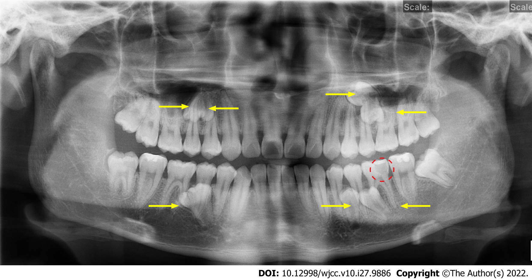
Figure 1 Preoperative panoramic radiograph.
The yellow arrows show the positions of the seven impacted SNTs. The red circle shows a local low-density area close to the pulp cavity in tooth #36.
- Citation: Wang Z, Zhao SY, He WS, Yu F, Shi SJ, Xia XL, Luo XX, Xiao YH. Application of digital positioning guide plates for the surgical extraction of multiple impacted supernumerary teeth: A case report and review of literature. World J Clin Cases 2022; 10(27): 9886-9896
- URL: https://www.wjgnet.com/2307-8960/full/v10/i27/9886.htm
- DOI: https://dx.doi.org/10.12998/wjcc.v10.i27.9886