
















©The Author(s) 2022.
World J Clin Cases. Sep 26, 2022; 10(27): 9760-9767
Published online Sep 26, 2022. doi: 10.12998/wjcc.v10.i27.9760
Published online Sep 26, 2022. doi: 10.12998/wjcc.v10.i27.9760
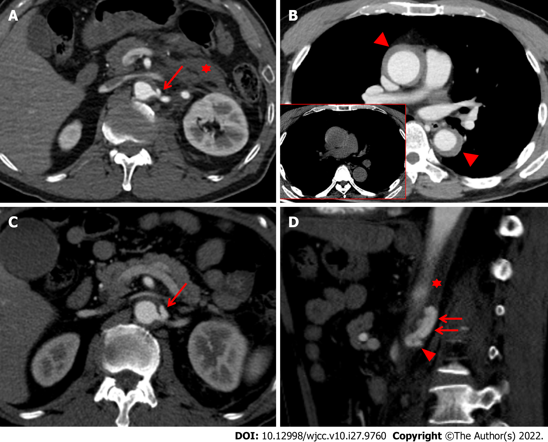
Figure 1 Initial computed tomography scans taken 1 h (A and B) after injury, and follow-up CT scans taken 15 d (C and D) after injury.
A: The initial abdominal computed tomography (CT) scan also revealed focal out-pouching enhancing nodular lesion, suggesting a pseudoaneurysm (red arrow) arising from the proximal portion of the left renal artery with retroperitoneal hematoma (red asterisk); B: The initial chest CT scan taken one hour after injury showed Stanford type A intramural hematoma (IMH) (red arrowheads), which represent slightly hyperdensity on precontrast image (figure in red-border box); C: The CT scan reveals an increase in the size and extent of the pseudoaneurysm (red arrow) on the axial image; D: The pseudoaneurysm arising from the proximal renal artery (red arrowhead) was localized within aortic media (red asterisk), resulting in an intramural blood collection (red arrows) seen on the sagittal image.
- Citation: Kim Y, Lee JY, Lee JS, Ye JB, Kim SH, Sul YH, Yoon SY, Choi JH, Choi H. Endovascular treatment of traumatic renal artery pseudoaneurysm with a Stanford type A intramural haematoma: A case report. World J Clin Cases 2022; 10(27): 9760-9767
- URL: https://www.wjgnet.com/2307-8960/full/v10/i27/9760.htm
- DOI: https://dx.doi.org/10.12998/wjcc.v10.i27.9760