Copyright
©The Author(s) 2022.
World J Clin Cases. Sep 26, 2022; 10(27): 9670-9679
Published online Sep 26, 2022. doi: 10.12998/wjcc.v10.i27.9670
Published online Sep 26, 2022. doi: 10.12998/wjcc.v10.i27.9670
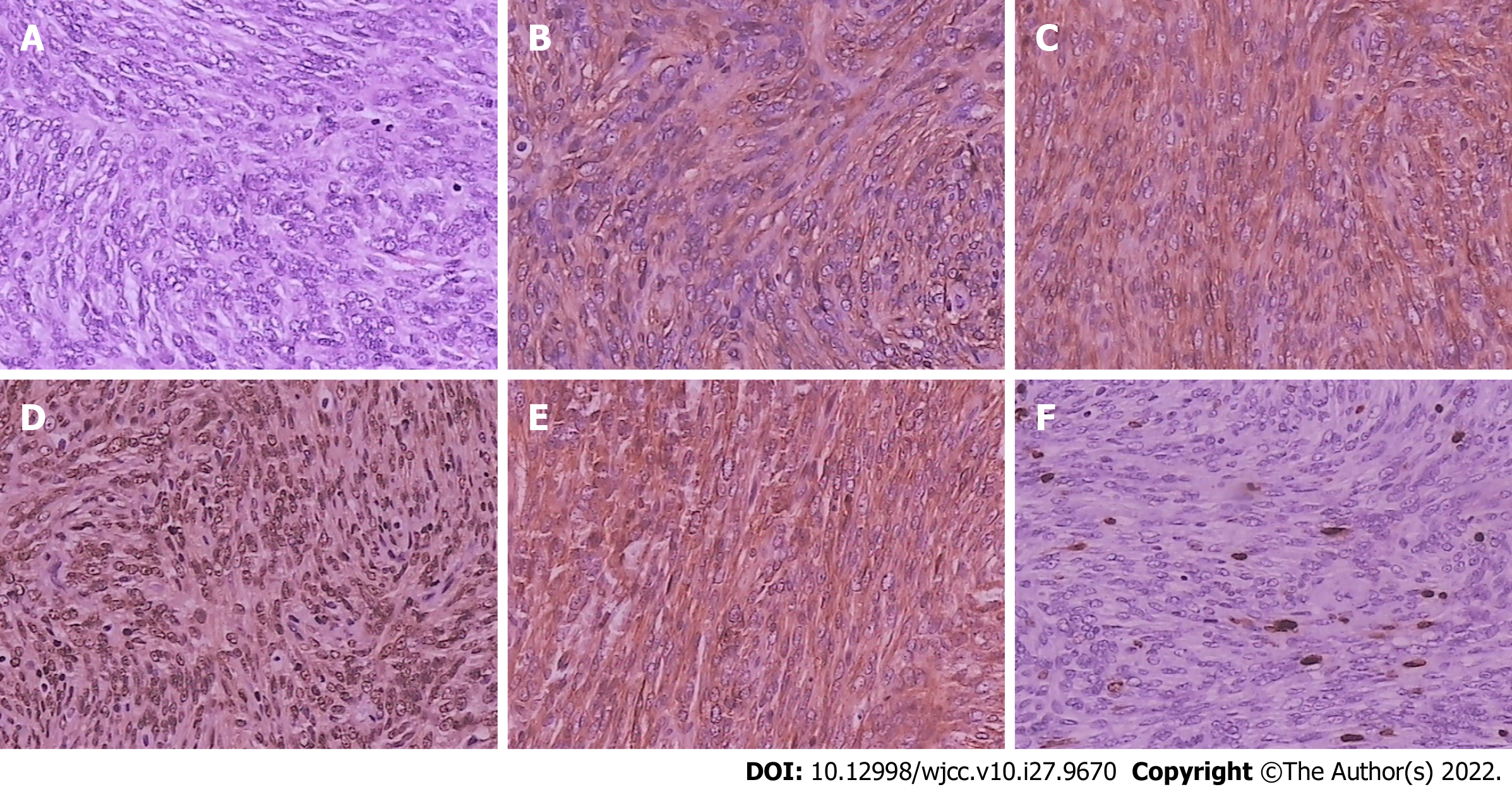
Figure 4 Histopathological and immunohistochemical examinations of tumors.
A: Tumor cells with oval nuclei, conspicuous nucleoli, and mitotic activity (hematoxylin and eosin staining, 200 ×); B: Tumor cells positive for CD34 (200 ×); C: Tumor cells positive for CD99 (200 ×); D: Tumor cells positive for STAT-6 (200 ×); E: Tumor cells positive for Bcl-2 (200 ×); F: Tumor cells positive for Ki-67 (close to 10%) (200 ×).
- Citation: Ren MY, Li J, Wu YX, Li RM, Zhang C, Liu LM, Wang JJ, Gao Y. Clinical characteristics and prognosis of orbital solitary fibrous tumor in patients from a Chinese tertiary eye hospital. World J Clin Cases 2022; 10(27): 9670-9679
- URL: https://www.wjgnet.com/2307-8960/full/v10/i27/9670.htm
- DOI: https://dx.doi.org/10.12998/wjcc.v10.i27.9670


