
















©The Author(s) 2022.
World J Clin Cases. Sep 26, 2022; 10(27): 9628-9640
Published online Sep 26, 2022. doi: 10.12998/wjcc.v10.i27.9628
Published online Sep 26, 2022. doi: 10.12998/wjcc.v10.i27.9628
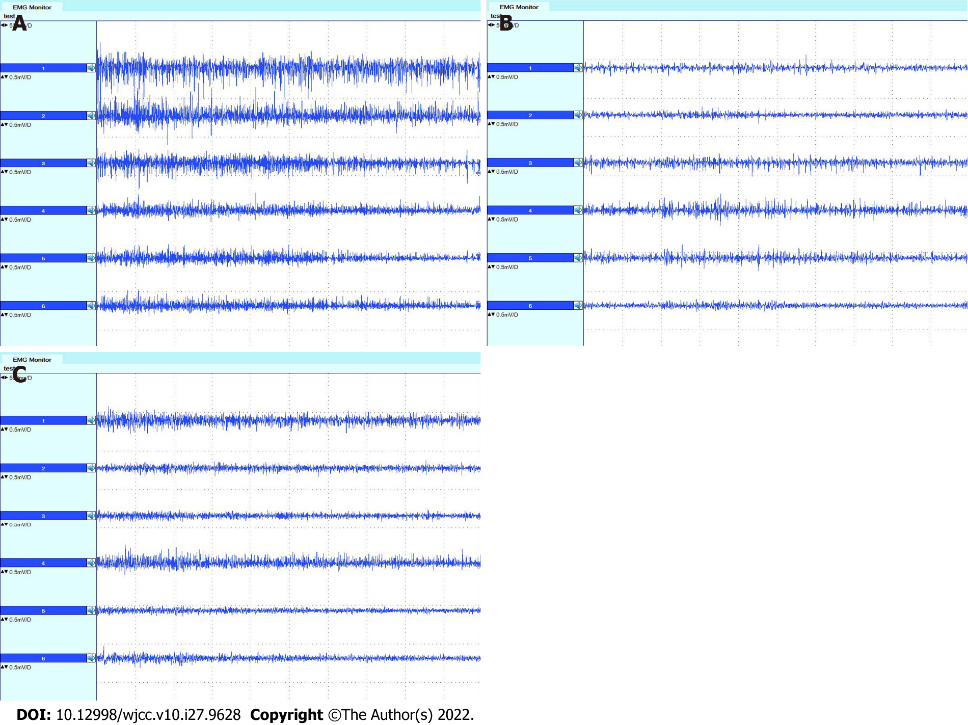
Figure 7 Electromyographs of the trapezius muscle.
A: Before surgery; B: Three months after surgery; C: Nine months after surgery. Electromyographs from top to bottom show the results of the descending portion on the left side, the descending portion on the right side, the horizontal portion on the left side, the horizontal portion on the right side, the ascending portion on the left side, and the horizontal portion on the right side, respectively.
- Citation: Wang JW, Zhang WB, Li F, Fang X, Yi ZQ, Xu XL, Peng X, Zhang WG. Anatomy and clinical application of suprascapular nerve to accessory nerve transfer. World J Clin Cases 2022; 10(27): 9628-9640
- URL: https://www.wjgnet.com/2307-8960/full/v10/i27/9628.htm
- DOI: https://dx.doi.org/10.12998/wjcc.v10.i27.9628