Copyright
©The Author(s) 2022.
World J Clin Cases. Sep 16, 2022; 10(26): 9510-9517
Published online Sep 16, 2022. doi: 10.12998/wjcc.v10.i26.9510
Published online Sep 16, 2022. doi: 10.12998/wjcc.v10.i26.9510
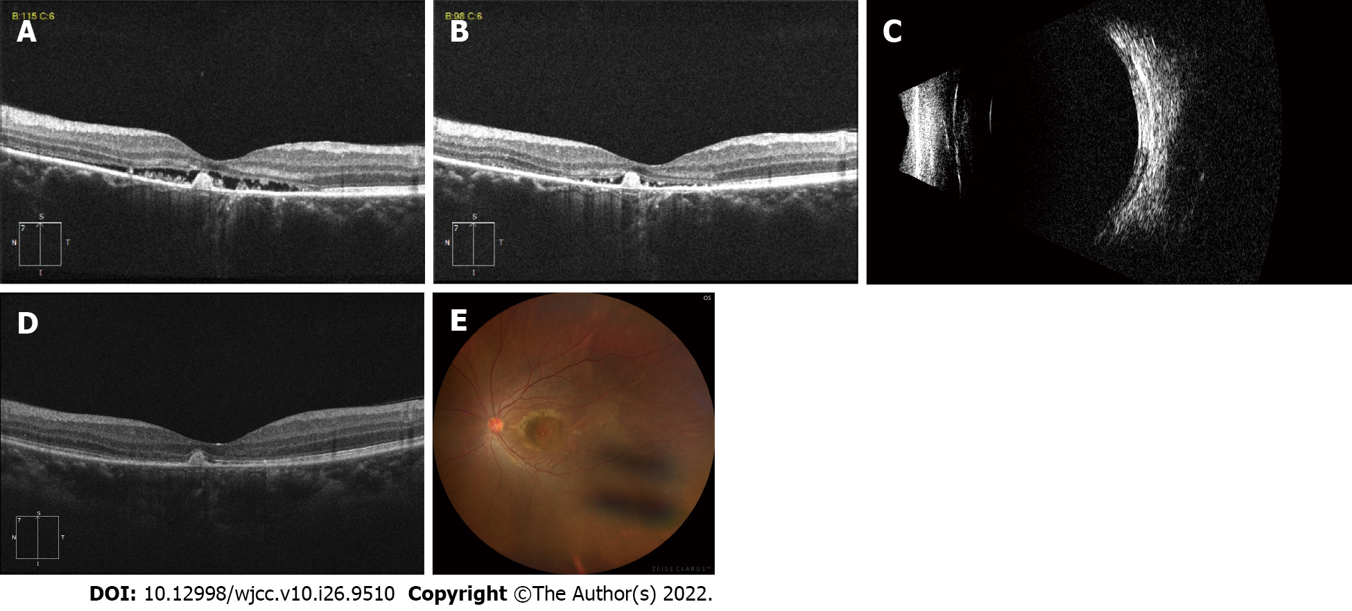
Figure 4 Examination images after the first and second intravitreal injections of conbercept.
A: Optical coherence tomography (OCT) showing a decrease in subretinal fluid with persistence of hyperreflective subretinal fibrin 1 mo after the first injection; B: OCT revealing a small amount of subretinal fluid with persistence of hyperreflective subretinal fibrin 1 mo after the second injection; C: B-scan ultrasonography revealing clear resolution of bullous retinal detachment 1 mo after the second injection; D: OCT revealing resolution of subretinal fluid with persistence of the hyperreflective subretinal fibrin 6 mo after the second intravitreal injection of conbercept; E: Fundus photograph of the left eye shows total resolution of the exudative detachment with subretinal exudation (fibrin) at the posterior pole 6 mo after the second intravitreal injection of conbercept.
- Citation: Xiang XL, Cao YH, Jiang TW, Huang ZR. Intravitreous injection of conbercept for bullous retinal detachment: A case report. World J Clin Cases 2022; 10(26): 9510-9517
- URL: https://www.wjgnet.com/2307-8960/full/v10/i26/9510.htm
- DOI: https://dx.doi.org/10.12998/wjcc.v10.i26.9510


