Copyright
©The Author(s) 2022.
World J Clin Cases. Sep 16, 2022; 10(26): 9510-9517
Published online Sep 16, 2022. doi: 10.12998/wjcc.v10.i26.9510
Published online Sep 16, 2022. doi: 10.12998/wjcc.v10.i26.9510
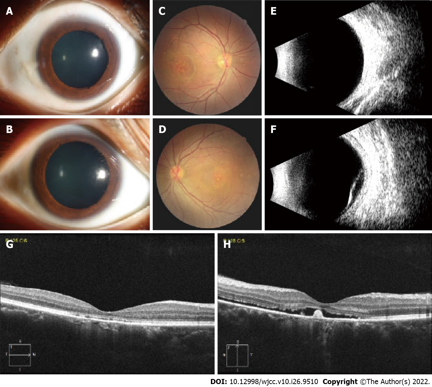
Figure 1 Anterior segment and fundus photograph, ophthalmic B-scan ultrasonography, and optical coherence tomography examination of the patient.
A: The anterior segment of the right eye is normal (dilated); B: The anterior segment of the left eye is normal (dilated); C: Fundus photograph shows white-yellow subretinal exudates in the posterior pole of the right eye; D: Fundus photograph of the left eye shows inferior exudative retinal detachment with subretinal exudation (fibrin) inferior to the macula; E: B-scan ultrasonography of the right eye is normal; F: B-scan ultrasonography confirms the bullous retinal detachment in the left eye; G: Optical coherence tomography (OCT) shows a discontinuous band of retinal pigment epithelium in the right eye; H: OCT shows neurosensory detachment at the fovea, with some hyperreflective material suggestive of fibrin in the left eye.
- Citation: Xiang XL, Cao YH, Jiang TW, Huang ZR. Intravitreous injection of conbercept for bullous retinal detachment: A case report. World J Clin Cases 2022; 10(26): 9510-9517
- URL: https://www.wjgnet.com/2307-8960/full/v10/i26/9510.htm
- DOI: https://dx.doi.org/10.12998/wjcc.v10.i26.9510


