Copyright
©The Author(s) 2022.
World J Clin Cases. Sep 6, 2022; 10(25): 9028-9035
Published online Sep 6, 2022. doi: 10.12998/wjcc.v10.i25.9028
Published online Sep 6, 2022. doi: 10.12998/wjcc.v10.i25.9028
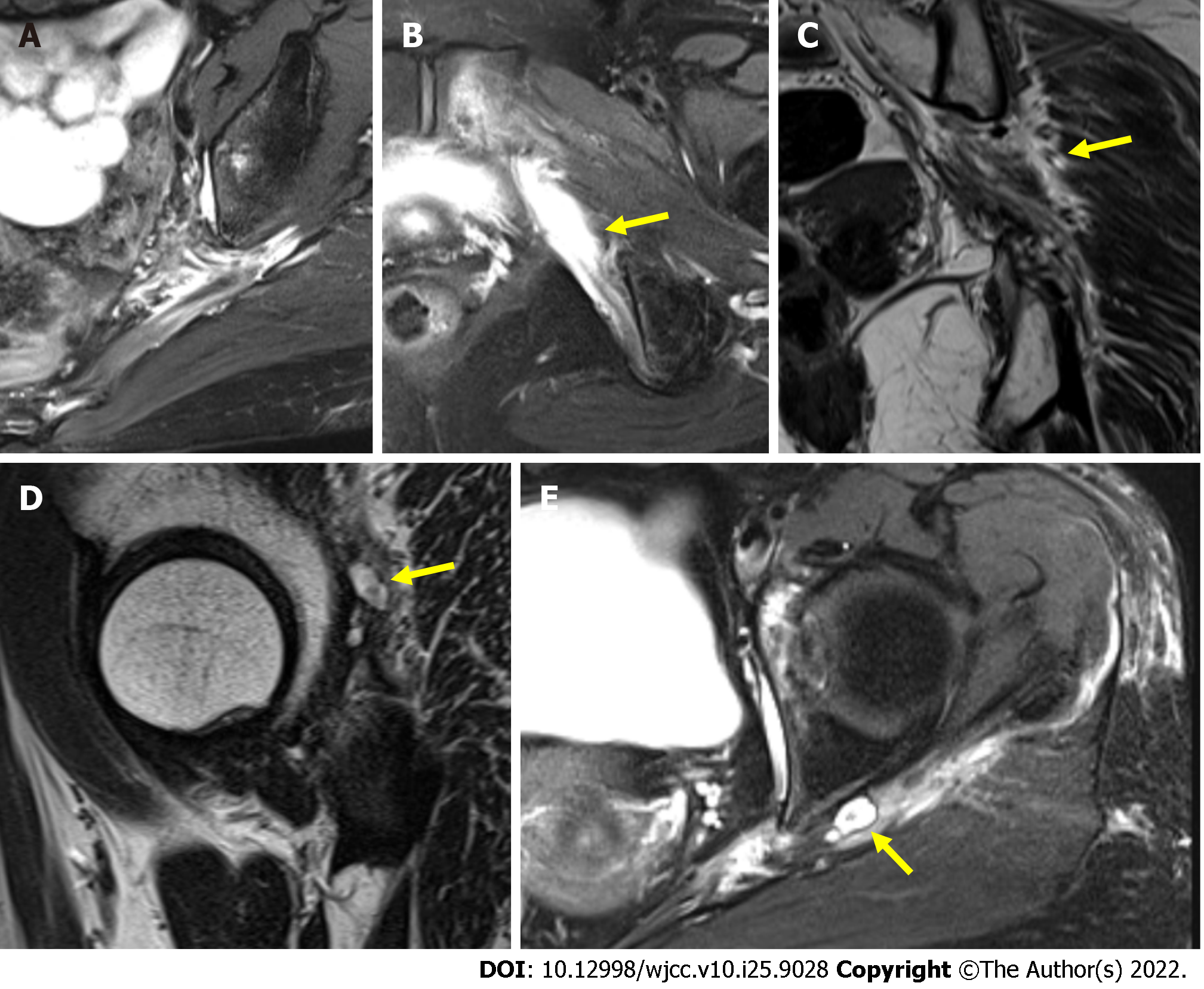
Figure 5 Postoperative T2-weighted Magnetic resonance image.
A: Magnetic resonance imaging finding shows that previously seen cystic mass is completely resected; B: Severely thickened left sciatic nerve is seen at right before greater sciatic foramen level and edematous change is accompanied around; C: Coronal view. Severe edema is around perifascicular fat which is covering nerve; D and E: Paralabral cyst. The cystic structure connected to hip joint with stalk stretch out to left ischial spine level. Yellow arrow indicates remnant cyst after excised multiple ganglionic masses in the deep gluteal space.
- Citation: Choi WK, Oh JS, Yoon SJ. Simultaneous laparoscopic and arthroscopic excision of a huge juxta-articular ganglionic cyst compressing the sciatic nerve: A case report. World J Clin Cases 2022; 10(25): 9028-9035
- URL: https://www.wjgnet.com/2307-8960/full/v10/i25/9028.htm
- DOI: https://dx.doi.org/10.12998/wjcc.v10.i25.9028


