Copyright
©The Author(s) 2022.
World J Clin Cases. Sep 6, 2022; 10(25): 9028-9035
Published online Sep 6, 2022. doi: 10.12998/wjcc.v10.i25.9028
Published online Sep 6, 2022. doi: 10.12998/wjcc.v10.i25.9028
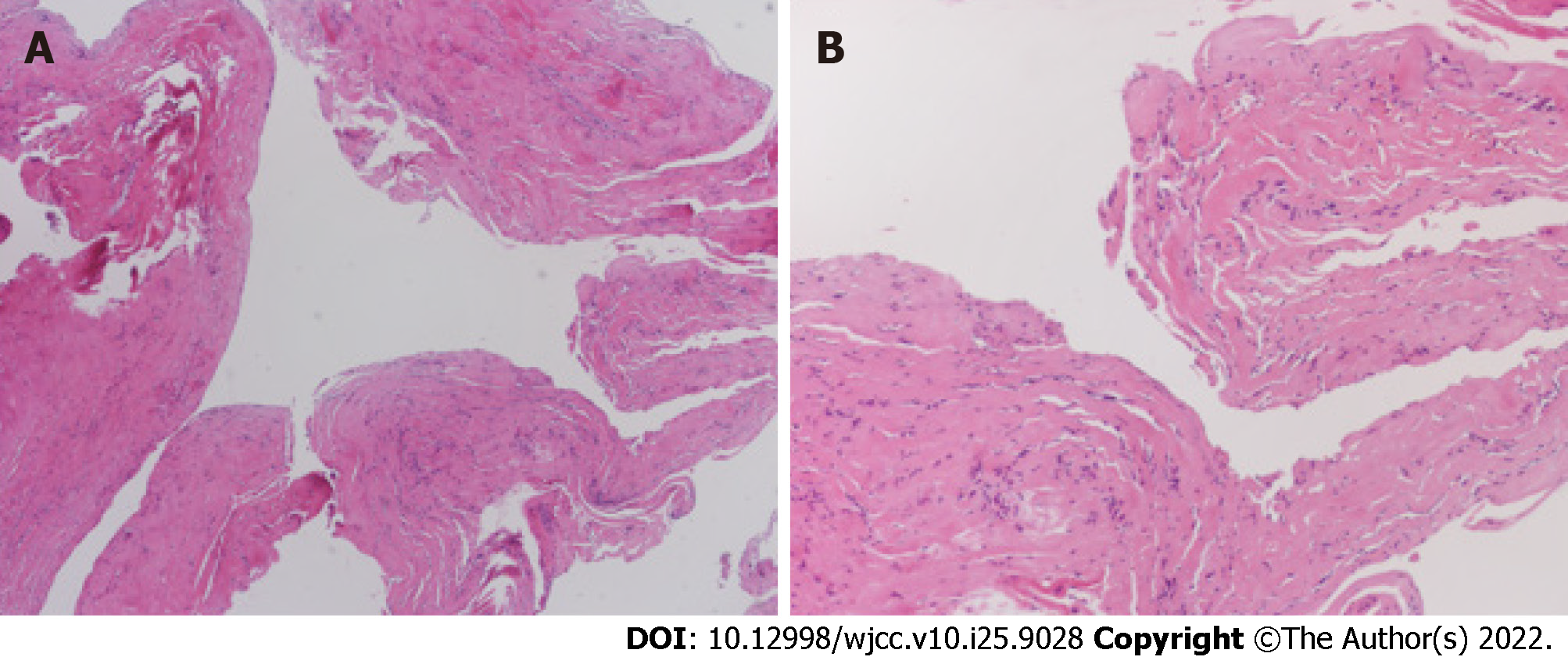
Figure 3 Histologic findings.
A: Microscopic examination showing cystically dilated fibrous tissues (hematoxylin and eosin stain, original magnification: × 40); B: The cyst wall is composed of fibroblasts, and the true epithelial lining is lacking (hematoxylin and eosin stain, original magnification: × 100).
- Citation: Choi WK, Oh JS, Yoon SJ. Simultaneous laparoscopic and arthroscopic excision of a huge juxta-articular ganglionic cyst compressing the sciatic nerve: A case report. World J Clin Cases 2022; 10(25): 9028-9035
- URL: https://www.wjgnet.com/2307-8960/full/v10/i25/9028.htm
- DOI: https://dx.doi.org/10.12998/wjcc.v10.i25.9028


