Copyright
©The Author(s) 2022.
World J Clin Cases. Sep 6, 2022; 10(25): 9028-9035
Published online Sep 6, 2022. doi: 10.12998/wjcc.v10.i25.9028
Published online Sep 6, 2022. doi: 10.12998/wjcc.v10.i25.9028
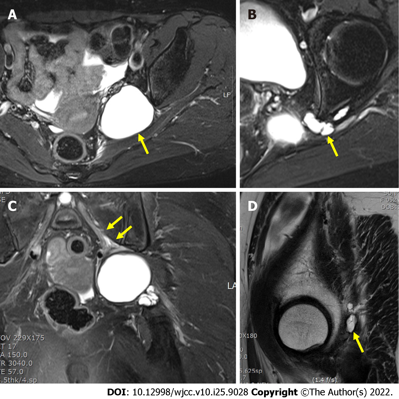
Figure 1 Preoperative T2-weighted Magnetic resonance image of the lesion.
The cystic lesion is seen as 5 cm × 5 cm × 4.6 cm sized with high signal intensity extending to greater sciatic notch. Stalk-like structure extended from left hip joint to main cyst lesion. A: TSE SPAIR transverse; B: TSE SPAIR transverse; C: TSE FS coronal; D: TSE sagittal.
- Citation: Choi WK, Oh JS, Yoon SJ. Simultaneous laparoscopic and arthroscopic excision of a huge juxta-articular ganglionic cyst compressing the sciatic nerve: A case report. World J Clin Cases 2022; 10(25): 9028-9035
- URL: https://www.wjgnet.com/2307-8960/full/v10/i25/9028.htm
- DOI: https://dx.doi.org/10.12998/wjcc.v10.i25.9028


