
















©The Author(s) 2022.
World J Clin Cases. Aug 26, 2022; 10(24): 8775-8781
Published online Aug 26, 2022. doi: 10.12998/wjcc.v10.i24.8775
Published online Aug 26, 2022. doi: 10.12998/wjcc.v10.i24.8775
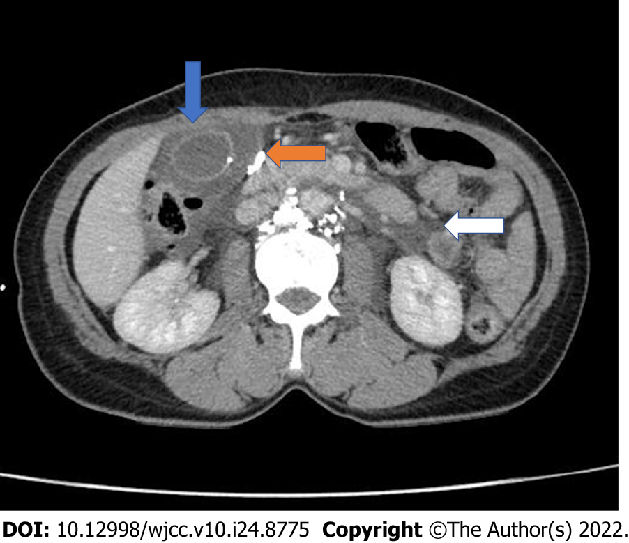
Figure 3 Contrast-enhanced computed tomography after intervention.
Axial image showed gallbladder wall thickening of approximately 14 mm and subserosal edema (blue arrow). Peripancreatic fluid is observed extending into the pararenal spaces (white arrow) and bilateral paracolic gutters. A small, high-density nodule is observed in the gallbladder wall (orange arrow).
- Citation: Dung LV, Hien MM, Tra My TT, Luu DT, Linh LT, Duc NM. Cholecystitis-an uncommon complication following thoracic duct embolization for chylothorax: A case report. World J Clin Cases 2022; 10(24): 8775-8781
- URL: https://www.wjgnet.com/2307-8960/full/v10/i24/8775.htm
- DOI: https://dx.doi.org/10.12998/wjcc.v10.i24.8775