Copyright
©The Author(s) 2022.
World J Clin Cases. Aug 26, 2022; 10(24): 8775-8781
Published online Aug 26, 2022. doi: 10.12998/wjcc.v10.i24.8775
Published online Aug 26, 2022. doi: 10.12998/wjcc.v10.i24.8775
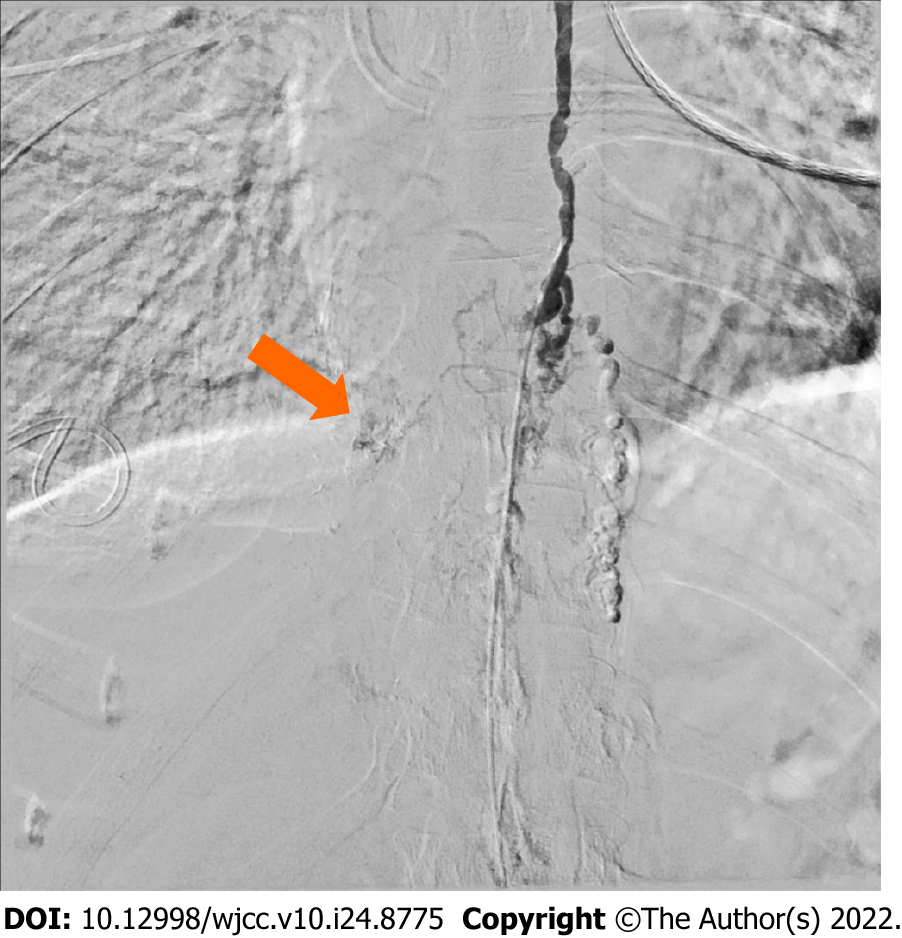
Figure 2 Intranodal lymphangiogram using digital subtraction angiography prior to treatment.
Frontal image of digital subtraction angiography revealed dilated lymphatic vessels and a lymphatic malformation in the lower thoracic duct (orange arrow), resulting in the leakage of contrast agent into the right pleural space.
- Citation: Dung LV, Hien MM, Tra My TT, Luu DT, Linh LT, Duc NM. Cholecystitis-an uncommon complication following thoracic duct embolization for chylothorax: A case report. World J Clin Cases 2022; 10(24): 8775-8781
- URL: https://www.wjgnet.com/2307-8960/full/v10/i24/8775.htm
- DOI: https://dx.doi.org/10.12998/wjcc.v10.i24.8775


