Copyright
©The Author(s) 2022.
World J Clin Cases. Aug 26, 2022; 10(24): 8679-8685
Published online Aug 26, 2022. doi: 10.12998/wjcc.v10.i24.8679
Published online Aug 26, 2022. doi: 10.12998/wjcc.v10.i24.8679
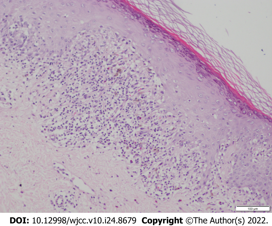
Figure 4 Skin histopathology (hematoxylin-eosin staining, 100×).
Histopathological examination showed reticular hyperkeratosis of the stratum corneum, wedge-shaped thickening of granular layer, irregular thickening of spinous layer, basal cell vacuolization and liquefaction, compact bandlike lymphocytic infiltration in superficial dermis, sporadic infiltration of chromatophilic cells, which shows typical features of lichen planus.
- Citation: Dong S, Zhu WJ, Xu M, Zhao XQ, Mou Y. Unilateral lichen planus with Blaschko line distribution: A case report. World J Clin Cases 2022; 10(24): 8679-8685
- URL: https://www.wjgnet.com/2307-8960/full/v10/i24/8679.htm
- DOI: https://dx.doi.org/10.12998/wjcc.v10.i24.8679


