Copyright
©The Author(s) 2022.
World J Clin Cases. Aug 26, 2022; 10(24): 8679-8685
Published online Aug 26, 2022. doi: 10.12998/wjcc.v10.i24.8679
Published online Aug 26, 2022. doi: 10.12998/wjcc.v10.i24.8679
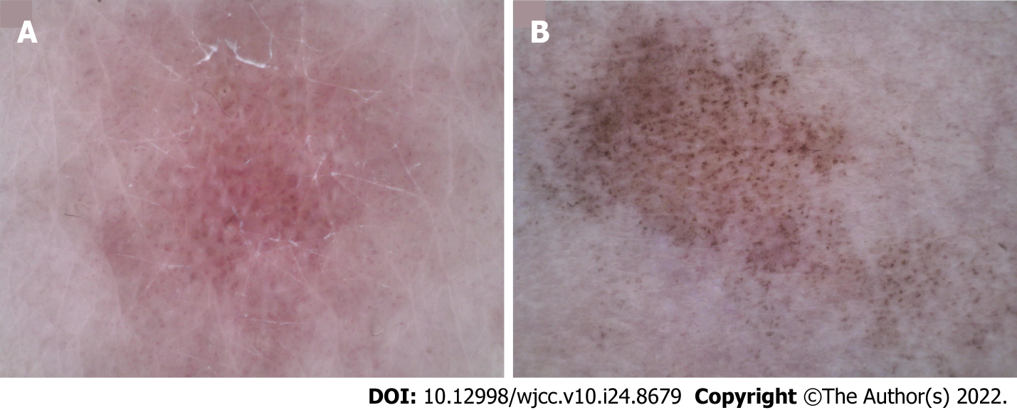
Figure 3 Dermoscopic photographs (50×).
A: Before treatment, linear and punctured vessels were seen under dermoscopy. The vascular structure was arranged radially with obvious white stripes; B: After treatment, the vascular structure disappeared, leaving blue-gray spots and faint white reticular stripes.
- Citation: Dong S, Zhu WJ, Xu M, Zhao XQ, Mou Y. Unilateral lichen planus with Blaschko line distribution: A case report. World J Clin Cases 2022; 10(24): 8679-8685
- URL: https://www.wjgnet.com/2307-8960/full/v10/i24/8679.htm
- DOI: https://dx.doi.org/10.12998/wjcc.v10.i24.8679


