Copyright
©The Author(s) 2022.
World J Clin Cases. Aug 16, 2022; 10(23): 8406-8416
Published online Aug 16, 2022. doi: 10.12998/wjcc.v10.i23.8406
Published online Aug 16, 2022. doi: 10.12998/wjcc.v10.i23.8406
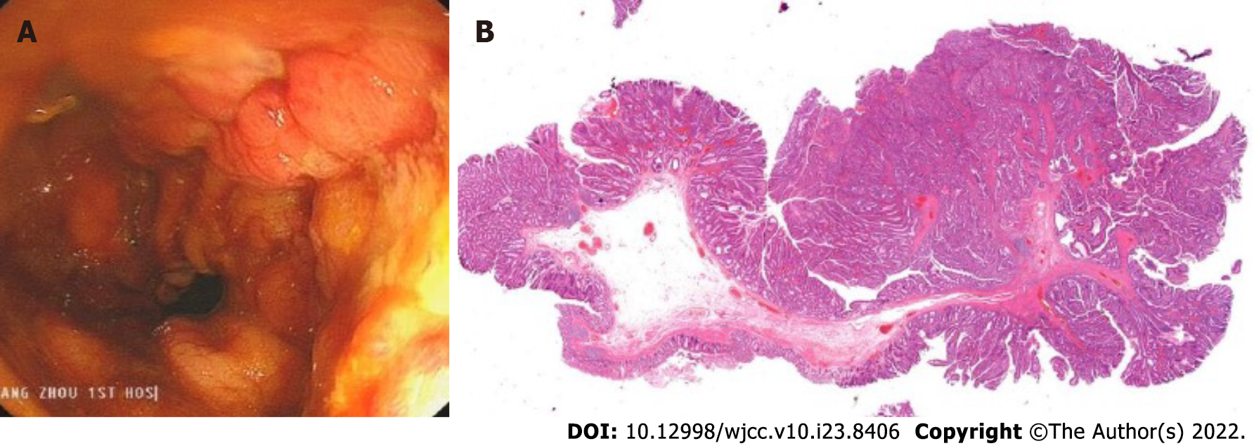
Figure 6 Removal of the metal stent and pathological findings.
A: After the metal stent was removed, it was observed that the wound began to heal, and the perforation and significant injury were no longer present; B: Tubular-villous adenoma with part of high-grade intraepithelial neoplasia (intramucosal carcinoma), infiltrating the muscularis mucosae, and with clean margin.
- Citation: Cheng SL, Xie L, Wu HW, Zhang XF, Lou LL, Shen HZ. Metal stent combined with ileus drainage tube for the treatment of delayed rectal perforation: A case report. World J Clin Cases 2022; 10(23): 8406-8416
- URL: https://www.wjgnet.com/2307-8960/full/v10/i23/8406.htm
- DOI: https://dx.doi.org/10.12998/wjcc.v10.i23.8406


