Copyright
©The Author(s) 2022.
World J Clin Cases. Aug 16, 2022; 10(23): 8392-8399
Published online Aug 16, 2022. doi: 10.12998/wjcc.v10.i23.8392
Published online Aug 16, 2022. doi: 10.12998/wjcc.v10.i23.8392
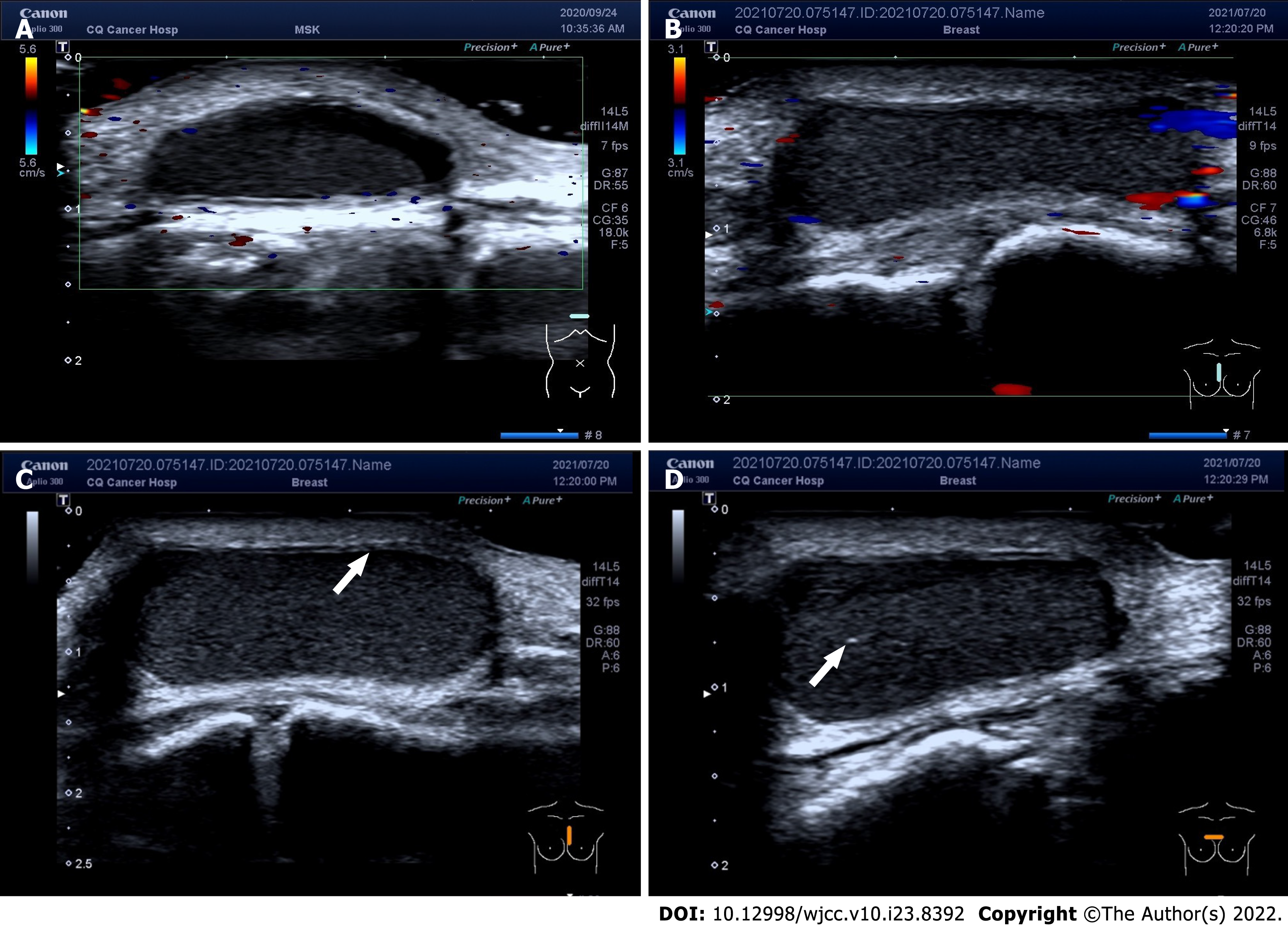
Figure 2 Imaging prior to surgery.
A: A low-to-no-echo area of about 8 mm × 19 mm × 26 mm in the patient’s subcutaneous soft tissue. The boundaries of the area were clear, and the morphology was clear. Furthermore, this area showed no obvious blood flow signal (September 24, 2020); B: A low-echo nodule of about 9 mm × 21 mm × 26 mm in the patient’s subcutaneous soft tissue. The boundaries of the nodule were clear, and the morphology was clear. At the same time, no intact envelope was found. A dense dot weak echo was found inside. The internal blood flow signal was not intense (July 20, 2021); C: Most of the cyst borders did not resemble the smooth walls of typical cysts, and the cyst appeared as a frizzy capsule instead. Echoes of the smooth wall can be seen in a few areas (white arrow; July 20, 2021); D: The inside of the cyst showed a relatively homogeneous low echo, and a small amount of oxalate calcification (white arrow) was found in some areas. The inflammation was mainly manifested in the cyst wall (July 20, 2021).
- Citation: Ma B, Fu KW, Xie XD, Cheng Y, Wang SQ. Bronchogenic cysts with infection in the chest wall skin of a 64-year-old asymptomatic patient: A case report. World J Clin Cases 2022; 10(23): 8392-8399
- URL: https://www.wjgnet.com/2307-8960/full/v10/i23/8392.htm
- DOI: https://dx.doi.org/10.12998/wjcc.v10.i23.8392


