Copyright
©The Author(s) 2022.
World J Clin Cases. Aug 16, 2022; 10(23): 8375-8383
Published online Aug 16, 2022. doi: 10.12998/wjcc.v10.i23.8375
Published online Aug 16, 2022. doi: 10.12998/wjcc.v10.i23.8375
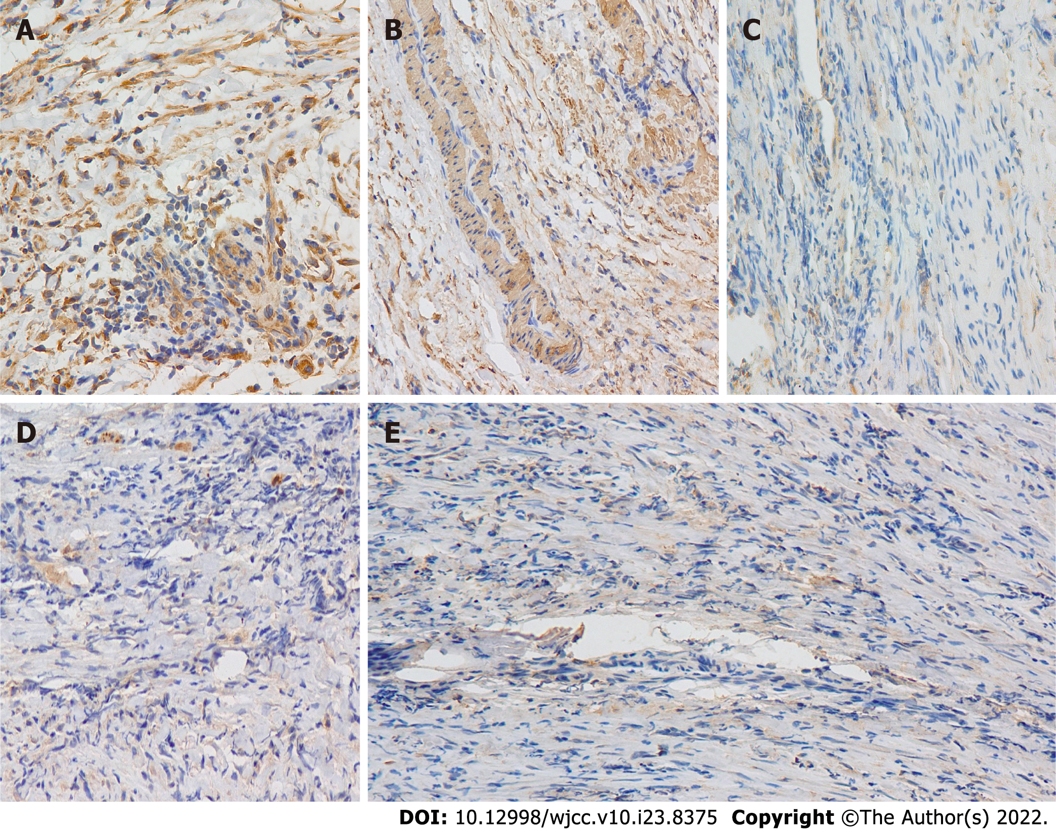
Figure 3 Photomicrograph.
A and B: Photomicrograph of inflammatory myofibroblastic tumor showing immunohistochemical positive for vimentin and smooth muscle actin (A: Original magnification: 400 ×; scale bar: 100 μm; and B: Original magnification: 200 ×; scale bar: 100 μm); C-E: Photomicrograph of inflammatory myofibroblastic tumor showing negative for desmin, S100 and ALK1 (Original magnification: 200 ×; scale bar: 100 μm) (Smooth muscle actin smooth muscle actin).
- Citation: Huang Y, Shu SN, Zhou H, Liu LL, Fang F. Infant biliary cirrhosis secondary to a biliary inflammatory myofibroblastic tumor: A case report and review of literature. World J Clin Cases 2022; 10(23): 8375-8383
- URL: https://www.wjgnet.com/2307-8960/full/v10/i23/8375.htm
- DOI: https://dx.doi.org/10.12998/wjcc.v10.i23.8375


