Copyright
©The Author(s) 2022.
World J Clin Cases. Aug 16, 2022; 10(23): 8352-8359
Published online Aug 16, 2022. doi: 10.12998/wjcc.v10.i23.8352
Published online Aug 16, 2022. doi: 10.12998/wjcc.v10.i23.8352
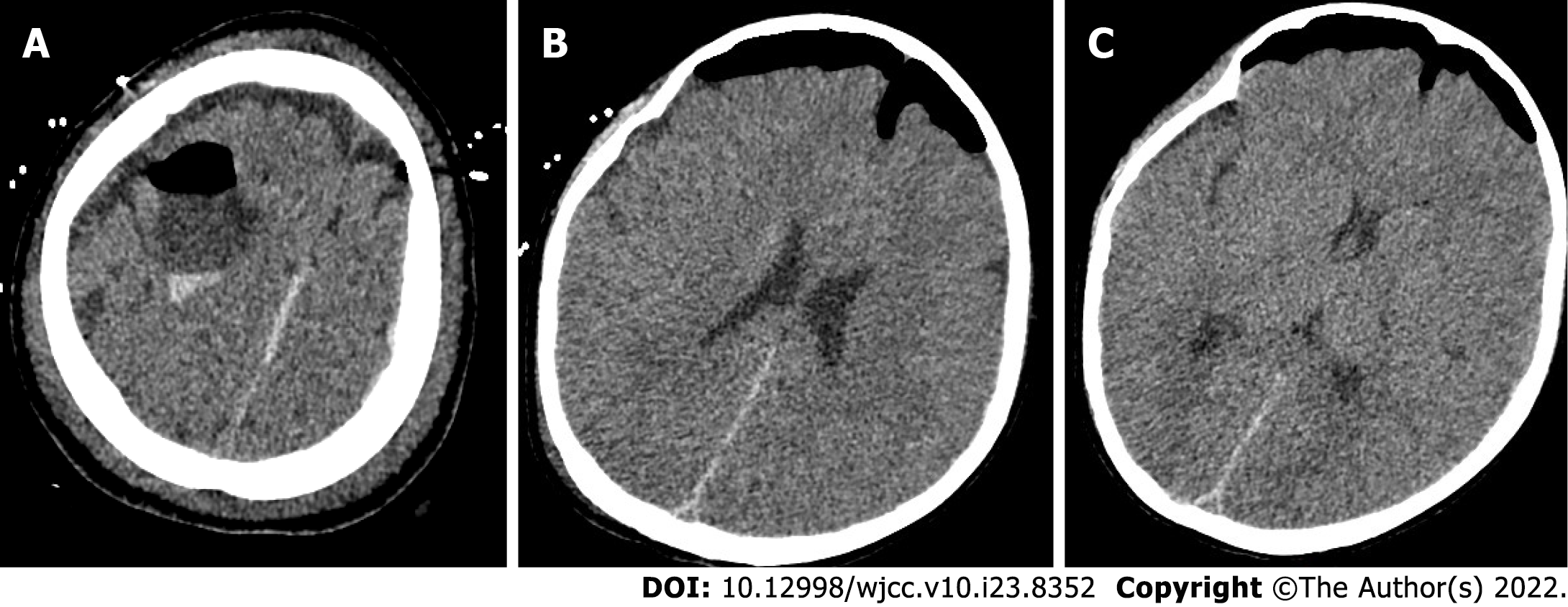
Figure 4 Postoperative head computed tomography.
A: The local bone of the right frontal area showed postoperative changes; B: The pressure in the arachnoid cyst was released; C: Right lateral ventricle and the brain tissue that was compressed and deformed before surgery gradually rebounded.
- Citation: Li WC, Li ML, Ding JW, Wang L, Wang SR, Wang YY, Xiao LF, Sun T. Incontinentia pigmenti with intracranial arachnoid cyst: A case report. World J Clin Cases 2022; 10(23): 8352-8359
- URL: https://www.wjgnet.com/2307-8960/full/v10/i23/8352.htm
- DOI: https://dx.doi.org/10.12998/wjcc.v10.i23.8352


