Copyright
©The Author(s) 2022.
World J Clin Cases. Aug 16, 2022; 10(23): 8344-8351
Published online Aug 16, 2022. doi: 10.12998/wjcc.v10.i23.8344
Published online Aug 16, 2022. doi: 10.12998/wjcc.v10.i23.8344
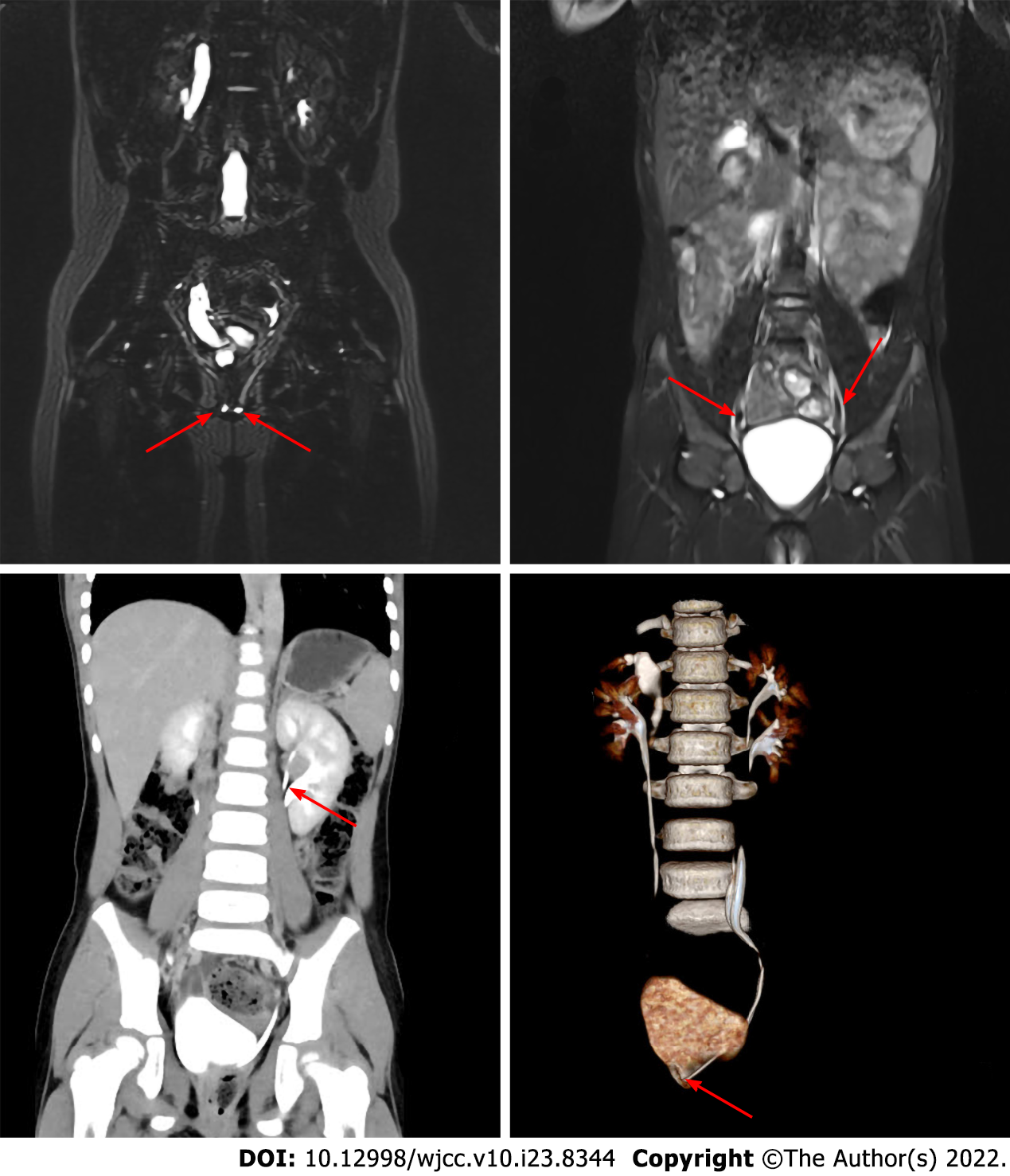
Figure 2 Imaging data.
A and B show abdomen magnetic resonance imaging, C and D show abdomen enhanced computed tomography. A: The arrow shows ectopic ureteral orifice; B: The arrow shows ureteral duplication; C: The arrow shows the duplication of the renal pelvis; D: The arrow shows the ectopic opening of the ureter.
- Citation: Wang SB, Wan L, Wang Y, Yi ZJ, Xiao C, Cao JZ, Liu XY, Tang RP, Luo Y. Laparoscopic treatment of bilateral duplex kidney and ectopic ureter: A case report. World J Clin Cases 2022; 10(23): 8344-8351
- URL: https://www.wjgnet.com/2307-8960/full/v10/i23/8344.htm
- DOI: https://dx.doi.org/10.12998/wjcc.v10.i23.8344


