Copyright
©The Author(s) 2022.
World J Clin Cases. Aug 16, 2022; 10(23): 8277-8283
Published online Aug 16, 2022. doi: 10.12998/wjcc.v10.i23.8277
Published online Aug 16, 2022. doi: 10.12998/wjcc.v10.i23.8277
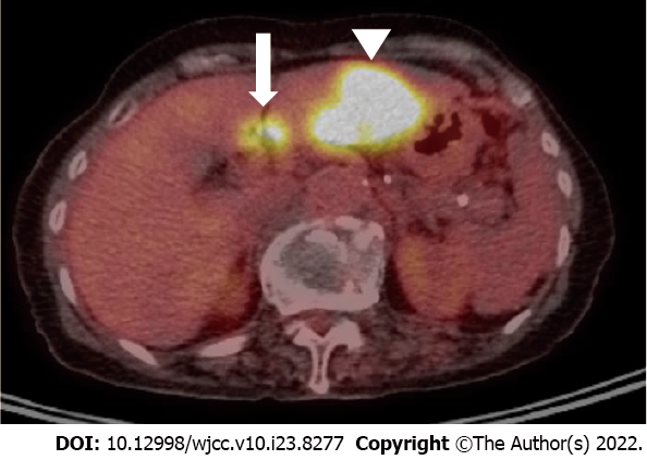
Figure 3 18F-fluorodeoxyglucose positron emission tomography-computed tomography examination.
Positron emission tomography-computed tomography examination image demonstrates a 4.5-cm hypermetabolic mass (arrowhead) in S3 and a 1.3-cm metastatic lymph with avid FDG uptake (arrow) in the node along the common hepatic artery.
- Citation: Noh BG, Seo HI, Park YM, Kim S, Hong SB, Lee SJ. Complete resection of large-cell neuroendocrine and hepatocellular carcinoma of the liver: A case report. World J Clin Cases 2022; 10(23): 8277-8283
- URL: https://www.wjgnet.com/2307-8960/full/v10/i23/8277.htm
- DOI: https://dx.doi.org/10.12998/wjcc.v10.i23.8277


