
















©The Author(s) 2022.
World J Clin Cases. Aug 16, 2022; 10(23): 8277-8283
Published online Aug 16, 2022. doi: 10.12998/wjcc.v10.i23.8277
Published online Aug 16, 2022. doi: 10.12998/wjcc.v10.i23.8277
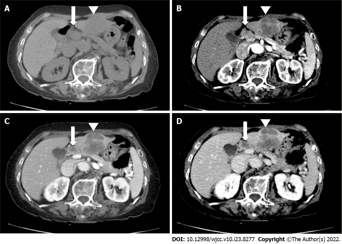
Figure 1 Abdominal contrast-enhanced computed tomography (CT) examination of the collision tumor.
A: Before contrast; B: Arterial phase; C: Portal venous phase; D: Delayed phases. Computed tomography (CT) images demonstrate a 4.5-cm mass (arrowhead) in S3. This mass shows peripheral rim enhancement during the arterial phase and washout during the portal venous phase and delayed phase. This mass was categorized using liver imaging reporting and data system M observations. CT images also demonstrate a 1.3-cm metastatic lymph (arrow) node along the common hepatic artery.
- Citation: Noh BG, Seo HI, Park YM, Kim S, Hong SB, Lee SJ. Complete resection of large-cell neuroendocrine and hepatocellular carcinoma of the liver: A case report. World J Clin Cases 2022; 10(23): 8277-8283
- URL: https://www.wjgnet.com/2307-8960/full/v10/i23/8277.htm
- DOI: https://dx.doi.org/10.12998/wjcc.v10.i23.8277