Copyright
©The Author(s) 2022.
World J Clin Cases. Aug 6, 2022; 10(22): 8045-8053
Published online Aug 6, 2022. doi: 10.12998/wjcc.v10.i22.8045
Published online Aug 6, 2022. doi: 10.12998/wjcc.v10.i22.8045
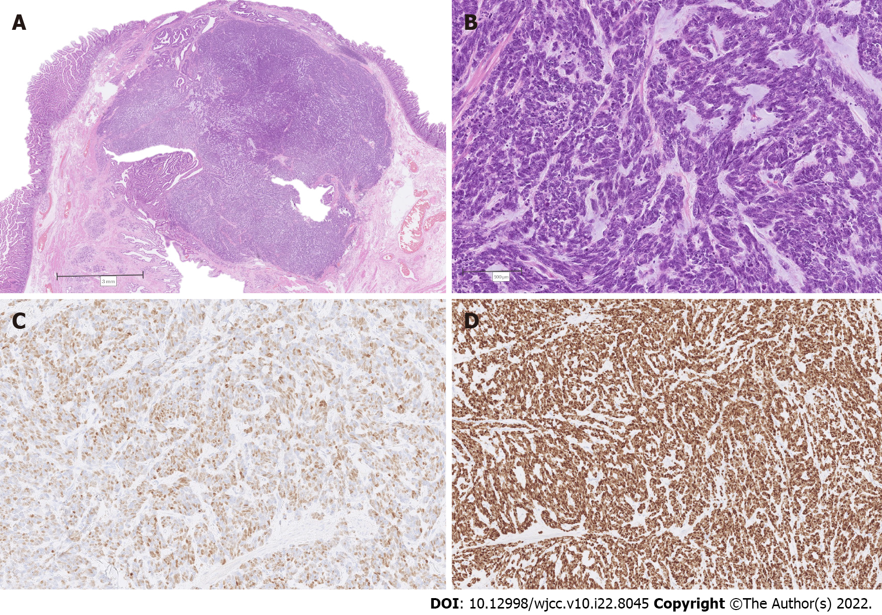
Figure 3 Combined intra-ampullary papillary-tubular neoplasm and neuroendocrine carcinoma.
A: Low power magnification depicting abrupt transition from the papillary tumor (upper and middle left) to solid nodular proliferation (middle and right); B: Higher magnification of the solid component, consistent with poorly differentiated neuroendocrine carcinoma; C: Immunohistochemistry for insulinoma-associated protein 1 depicting diffuse nuclear positivity; D: Immunohistochemistry revealing a 100% Ki-67 index in tumor cells.
- Citation: Zavrtanik H, Luzar B, Tomažič A. Intra-ampullary papillary-tubular neoplasm combined with ampullary neuroendocrine carcinoma: A case report. World J Clin Cases 2022; 10(22): 8045-8053
- URL: https://www.wjgnet.com/2307-8960/full/v10/i22/8045.htm
- DOI: https://dx.doi.org/10.12998/wjcc.v10.i22.8045


