Copyright
©The Author(s) 2022.
World J Clin Cases. Aug 6, 2022; 10(22): 7899-7905
Published online Aug 6, 2022. doi: 10.12998/wjcc.v10.i22.7899
Published online Aug 6, 2022. doi: 10.12998/wjcc.v10.i22.7899
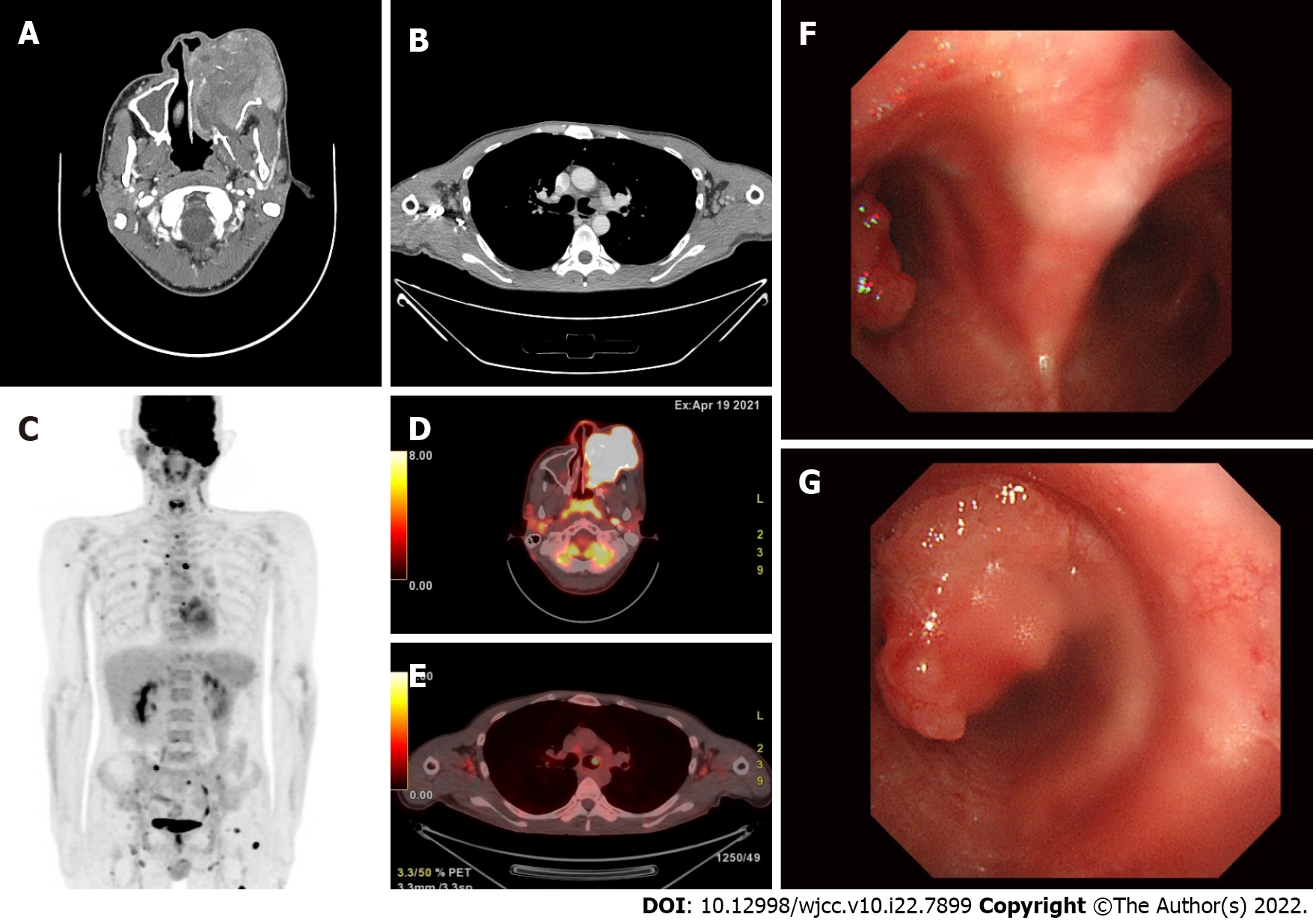
Figure 1 Imaging at admission.
A: Contrast-enhanced neck computed tomography shows a bulky mass in the left maxillary sinus extending to the orbit, nasal cavity, ethmoid sinus, infratemporal fossa, and pterygopalatine fossa; bone destruction extends to the nasal cavity; B: Contrast-enhanced chest computed tomography shows an enhanced nodule approximately 0.8 cm in size in the left main bronchus; C-E: 18F-fluorodeoxyglucose positron emission/computed tomography shows a large expansile hypermetabolic mass in the left maxillary sinus and hypermetabolic focal activity in the nasopharynx, multiple metastatic lymphadenopathies in both cervical and left supraclavicular areas, and multiple osseous metastases. There is a focal hypermetabolic nodular lesion in the left main bronchus; F and G: Bronchoscopy shows a 1.0-cm sized nodular lesion with pedicles arising from the anterior wall of the left main bronchus.
- Citation: Lee SB, Park CY, Lee HJ, Hong R, Kim WS, Park SG. Non-secretory multiple myeloma expressed as multiple extramedullary plasmacytoma with an endobronchial lesion mimicking metastatic cancer: A case report. World J Clin Cases 2022; 10(22): 7899-7905
- URL: https://www.wjgnet.com/2307-8960/full/v10/i22/7899.htm
- DOI: https://dx.doi.org/10.12998/wjcc.v10.i22.7899


