
















©The Author(s) 2022.
World J Clin Cases. Jul 26, 2022; 10(21): 7409-7414
Published online Jul 26, 2022. doi: 10.12998/wjcc.v10.i21.7409
Published online Jul 26, 2022. doi: 10.12998/wjcc.v10.i21.7409
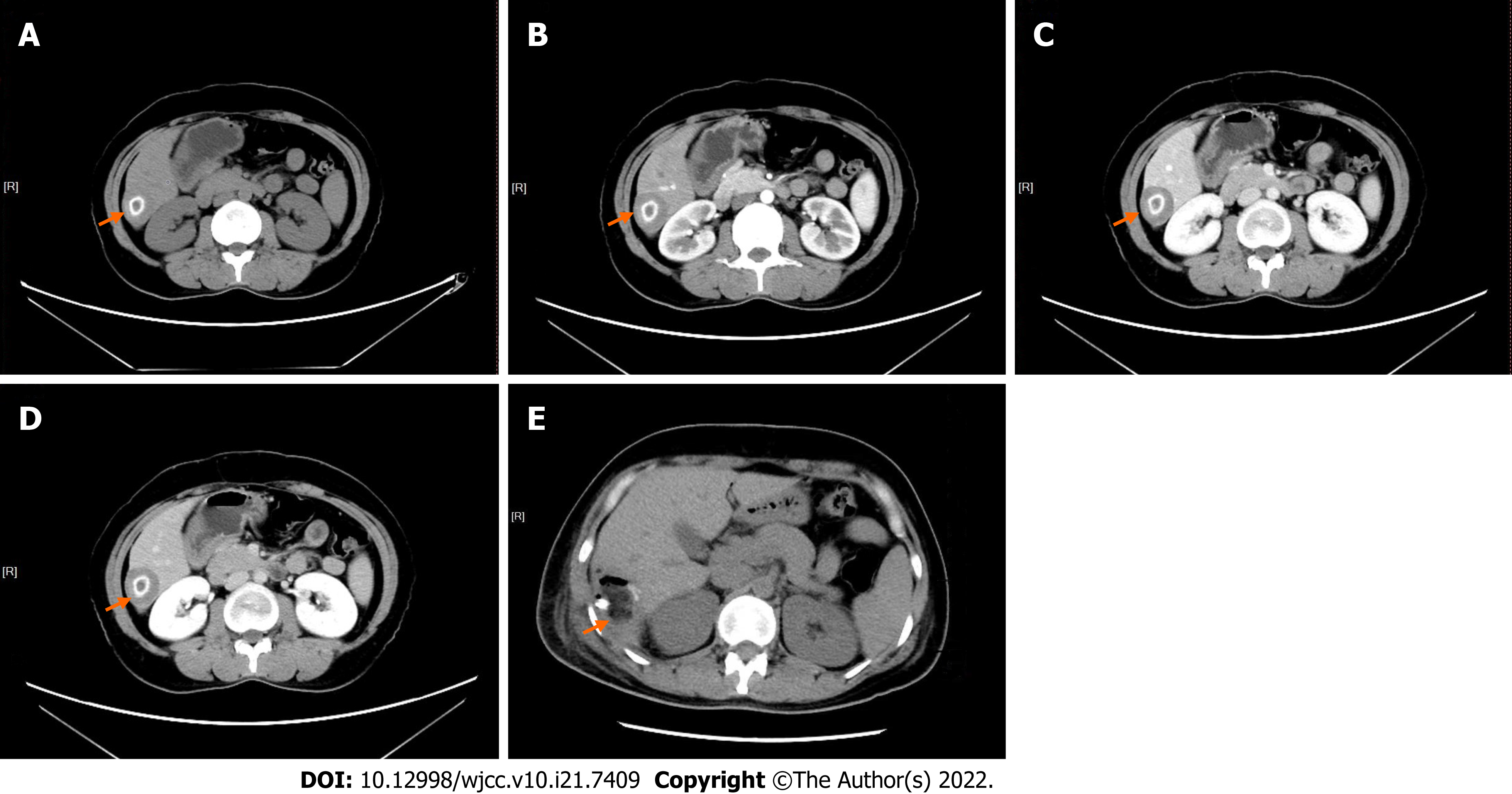
Figure 1 Computed tomography images of the lesion (orange arrow).
A: Plain scan image showing a slightly low density, round lesion and "ring"-like calcification in the lesion, measuring 3.4 cm × 2.7 cm; B: Arterial phase; C: Venous phase; D: Delayed phase; E: After the operation. The high signal in the surgical area was the manifestation of drainage tube, and the lesion had no enhancement in the arterial, venous, and delayed phases.
- Citation: Bao JP, Tian H, Wang HC, Wang CC, Li B. Solitary necrotic nodules of the liver with "ring"-like calcification: A case report. World J Clin Cases 2022; 10(21): 7409-7414
- URL: https://www.wjgnet.com/2307-8960/full/v10/i21/7409.htm
- DOI: https://dx.doi.org/10.12998/wjcc.v10.i21.7409