Copyright
©The Author(s) 2022.
World J Clin Cases. Jul 16, 2022; 10(20): 7138-7146
Published online Jul 16, 2022. doi: 10.12998/wjcc.v10.i20.7138
Published online Jul 16, 2022. doi: 10.12998/wjcc.v10.i20.7138
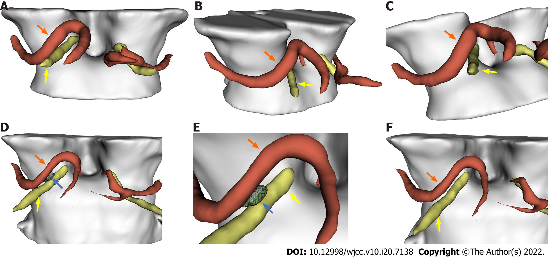
Figure 2 Representative three-dimensional images.
A-C: Demonstrated the right oculomotor nerve (ON) (yellow arrow) was compressed by the right posterior cerebral artery (PCA) (orange arrow) downwardly before surgery; D-F: Demonstrated the decompression of the right oculomotor nerve by the Telfon cottons (blue arrow) between the right PCA and the right oculomotor nerve 3 mo after surgery.
- Citation: Zhang J, Wei ZJ, Wang H, Yu YB, Sun HT. Microvascular decompression for a patient with oculomotor palsy caused by posterior cerebral artery compression: A case report and literature review . World J Clin Cases 2022; 10(20): 7138-7146
- URL: https://www.wjgnet.com/2307-8960/full/v10/i20/7138.htm
- DOI: https://dx.doi.org/10.12998/wjcc.v10.i20.7138


