Copyright
©The Author(s) 2022.
World J Clin Cases. Jul 16, 2022; 10(20): 7020-7028
Published online Jul 16, 2022. doi: 10.12998/wjcc.v10.i20.7020
Published online Jul 16, 2022. doi: 10.12998/wjcc.v10.i20.7020
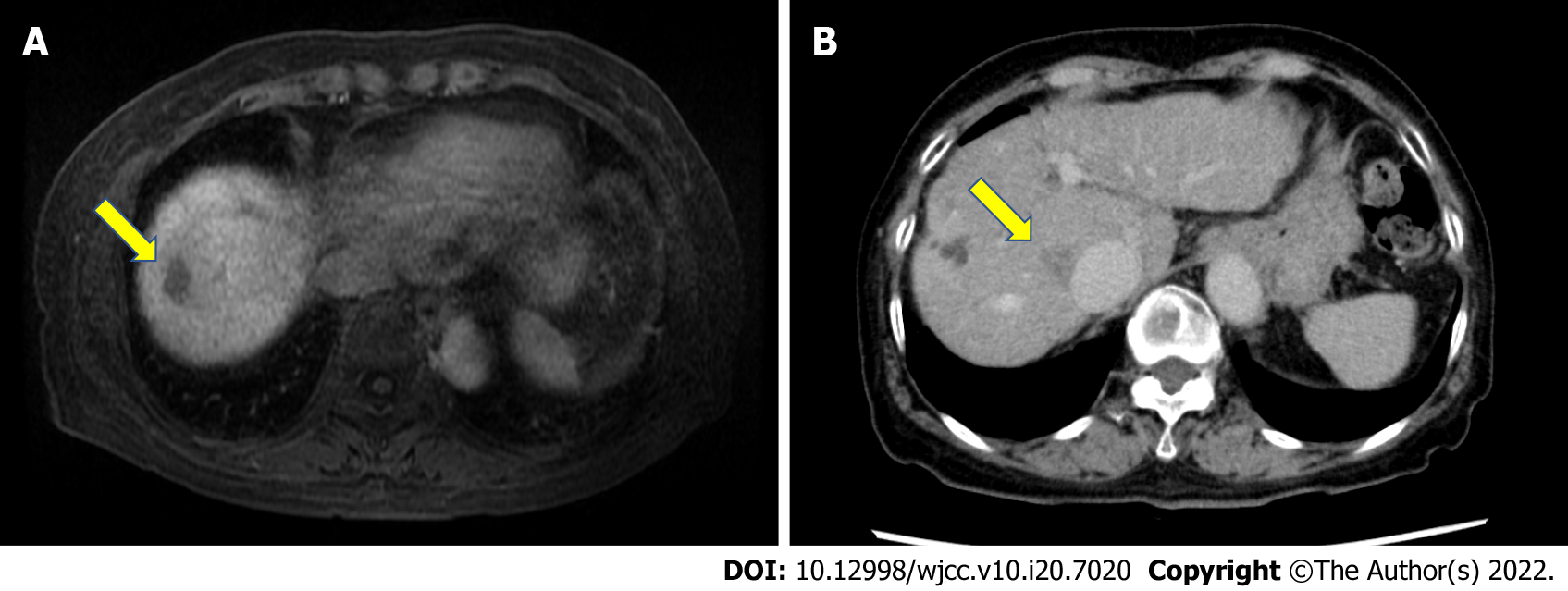
Figure 1 Location of tumors.
A: Gadolinium ethoxybenzyl diethylenetriamine pentaacetic acid-enhanced magnetic resonance imaging revealed a low-intensity area in segment VIII (S8) near the surface of the liver in the hepatobiliary phase (arrow); B: Abdominal contrast-enhanced computed tomography revealed a nodular lesion (20 mm) in S8 of the liver near the inferior vena cava, indicating washout in the delayed phase (arrow).
- Citation: Tsunoda J, Nishi T, Ito T, Inaguma G, Matsuzaki T, Seki H, Yasui N, Sakata M, Shimada A, Matsumoto H. Laparoscopic repair of diaphragmatic hernia associating with radiofrequency ablation for hepatocellular carcinoma: A case report. World J Clin Cases 2022; 10(20): 7020-7028
- URL: https://www.wjgnet.com/2307-8960/full/v10/i20/7020.htm
- DOI: https://dx.doi.org/10.12998/wjcc.v10.i20.7020


