Copyright
©The Author(s) 2022.
World J Clin Cases. Jul 16, 2022; 10(20): 7013-7019
Published online Jul 16, 2022. doi: 10.12998/wjcc.v10.i20.7013
Published online Jul 16, 2022. doi: 10.12998/wjcc.v10.i20.7013
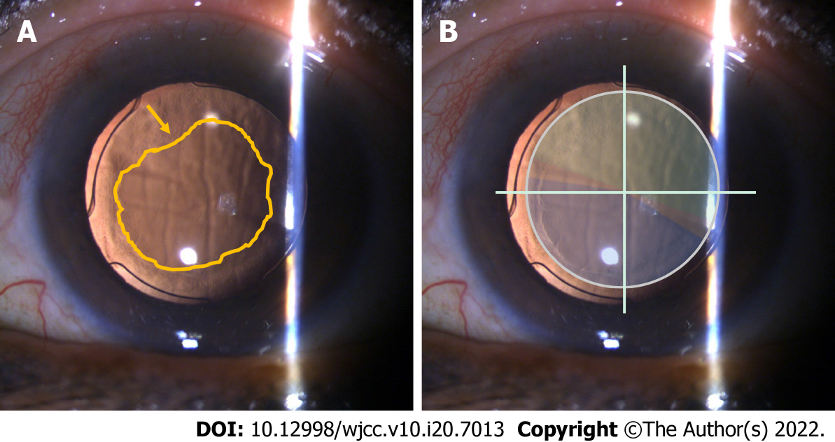
Figure 1 Slit lamp examinations 2 wk after phacoemulsification in the left eye.
A: The swelling of the corneal endothelium and the proliferation of lens epithelial cells are most pronounced (yellow arrow) over the surface of the intraocular lens; B: The intraocular lens is centered with the near segment placed inferonasally (blue: near segment; green: distant segment).
- Citation: Fan C, Zhou Y, Jiang J. Secondary positioning of rotationally asymmetric refractive multifocal intraocular lens in a patient with glaucoma: A case report. World J Clin Cases 2022; 10(20): 7013-7019
- URL: https://www.wjgnet.com/2307-8960/full/v10/i20/7013.htm
- DOI: https://dx.doi.org/10.12998/wjcc.v10.i20.7013


