Copyright
©The Author(s) 2022.
World J Clin Cases. Jul 16, 2022; 10(20): 6769-6783
Published online Jul 16, 2022. doi: 10.12998/wjcc.v10.i20.6769
Published online Jul 16, 2022. doi: 10.12998/wjcc.v10.i20.6769
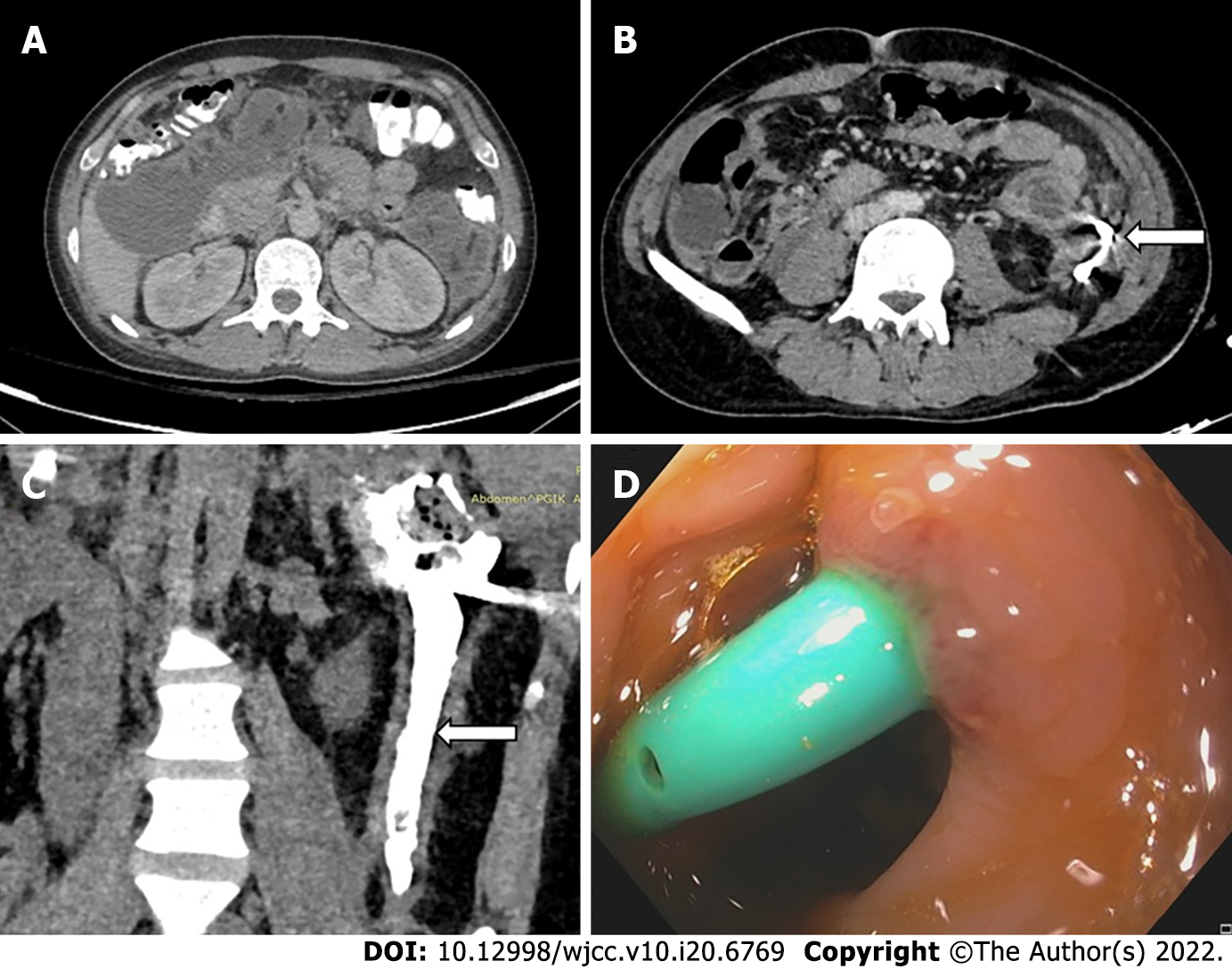
Figure 7 Gastrointestinal fistula formation after percutaneous catheter placement.
A: Pre percutaneous catheter (PCD) computed tomography (CT) showing walled off necrosis in left paracolic gutter (PCG) and subhepatic space; B: Axial CT showing pigtail in situ in the PCG collection (arrow); C: CT-PCD gram (coronal section) showing communication of the collection with descending colon with contrast in the lumen (arrow); D: Colonoscopic image showing pigtail tip in the colon lumen.
- Citation: Bansal A, Gupta P, Singh AK, Shah J, Samanta J, Mandavdhare HS, Sharma V, Sinha SK, Dutta U, Sandhu MS, Kochhar R. Drainage of pancreatic fluid collections in acute pancreatitis: A comprehensive overview. World J Clin Cases 2022; 10(20): 6769-6783
- URL: https://www.wjgnet.com/2307-8960/full/v10/i20/6769.htm
- DOI: https://dx.doi.org/10.12998/wjcc.v10.i20.6769


