Copyright
©The Author(s) 2022.
World J Clin Cases. Jan 14, 2022; 10(2): 709-716
Published online Jan 14, 2022. doi: 10.12998/wjcc.v10.i2.709
Published online Jan 14, 2022. doi: 10.12998/wjcc.v10.i2.709
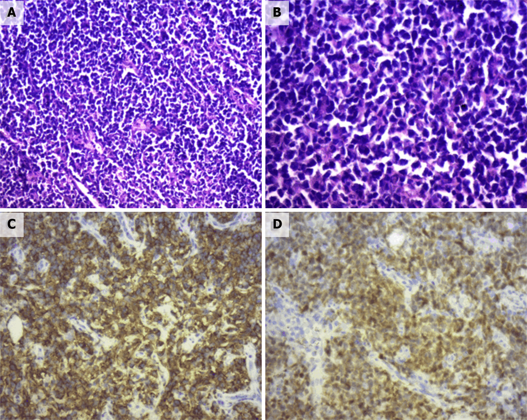
Figure 2 Pathological section.
A: Hematoxylin & eosin (HE, × 100). Diffuse infiltration and growth of tumor cells; B: HE (× 200). Tumor cells were composed of medium to large lymphoid cells, most of which were round and oval, double chromotropic or basophilic, containing less cytoplasm and large nuclei; C: Immunohistochemical SP method of CD20 staining. Strong positively stained cell membrane in all tumor cells (the brownish yellow part of the picture); D: Immunohistochemical SP method of CD20 staining. Strong positively stained cell membrane in all tumor cells (the brownish yellow part of the picture).
- Citation: Fan ZN, Shi HJ, Xiong BB, Zhang JS, Wang HF, Wang JS. Primary adrenal diffuse large B-cell lymphoma with normal adrenal cortex function: A case report. World J Clin Cases 2022; 10(2): 709-716
- URL: https://www.wjgnet.com/2307-8960/full/v10/i2/709.htm
- DOI: https://dx.doi.org/10.12998/wjcc.v10.i2.709


