Copyright
©The Author(s) 2022.
World J Clin Cases. Jan 14, 2022; 10(2): 685-690
Published online Jan 14, 2022. doi: 10.12998/wjcc.v10.i2.685
Published online Jan 14, 2022. doi: 10.12998/wjcc.v10.i2.685
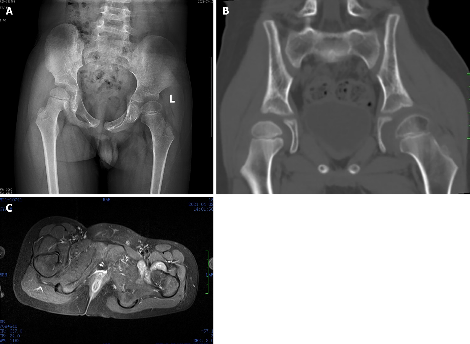
Figure 1 Radiology result of this patient.
A: X-ray shows no specific findings, and the left femoral head epiphysis was slightly flattened; B: Computed tomography shows decreased density of the femoral head epiphysis in the left hip joint and a widened gap in the left hip joint; C: Magnetic resonance imaging shows the synovium around the left hip joint of the child was thickened and a part of the synovium was nodular.
- Citation: Yi RB, Gong HL, Arthur DT, Wen J, Xiao S, Tang ZW, Xiang F, Wang KJ, Song ZQ. Synovial chondromatosis of the hip joint in a 6 year-old child: A case report. World J Clin Cases 2022; 10(2): 685-690
- URL: https://www.wjgnet.com/2307-8960/full/v10/i2/685.htm
- DOI: https://dx.doi.org/10.12998/wjcc.v10.i2.685


