Copyright
©The Author(s) 2022.
World J Clin Cases. Jul 6, 2022; 10(19): 6679-6687
Published online Jul 6, 2022. doi: 10.12998/wjcc.v10.i19.6679
Published online Jul 6, 2022. doi: 10.12998/wjcc.v10.i19.6679
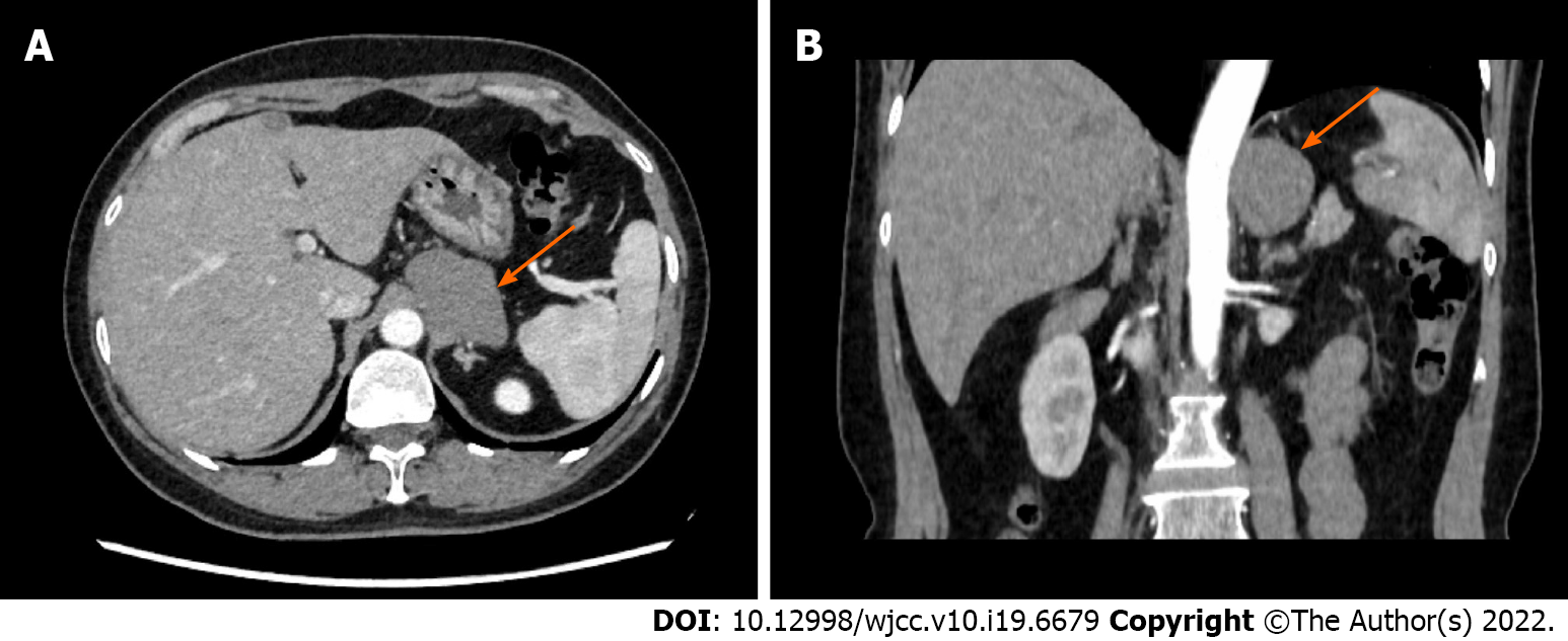
Figure 1 Abdominal enhanced computed tomography image.
A: Transverse section; B: Coronal section. The mass was located around the left adrenal gland. The 54 mm × 40 mm oval mass showed a clear boundary and a uniform density of 85 HU.
- Citation: Gong YY, Qian X, Liang B, Jiang MD, Liu J, Tao X, Luo J, Liu HJ, Feng YG. Retroperitoneal tumor finally diagnosed as a bronchogenic cyst: A case report and review of literature. World J Clin Cases 2022; 10(19): 6679-6687
- URL: https://www.wjgnet.com/2307-8960/full/v10/i19/6679.htm
- DOI: https://dx.doi.org/10.12998/wjcc.v10.i19.6679


