
















©The Author(s) 2022.
World J Clin Cases. Jul 6, 2022; 10(19): 6626-6635
Published online Jul 6, 2022. doi: 10.12998/wjcc.v10.i19.6626
Published online Jul 6, 2022. doi: 10.12998/wjcc.v10.i19.6626
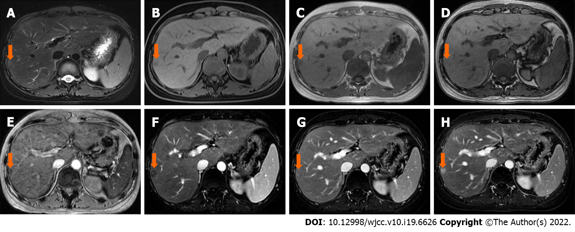
Figure 2 A 30-year-old female diagnosed with intrahepatic extramedullary hematopoiesis was confirmed by biopsy.
A and B: The lesion (arrow) located in the subcapsular of segment VI/VII was slightly hyperintense on T2 weighted image (WI)-fat saturation (FS) (A) and slightly hypointense on T1WI-FS (B); C-E: The lesion showed a lower signal intensity on the in-phase (C) than on the out-phase (D) image, and signal loss on susceptibility weighted imaging (E); F-H: In dynamic series, the lesion was mildly enhanced in the arterial phase (F), with areas of progressive and prolonged enhancement in the portal venous (G) and delayed phases (H).
- Citation: Luo M, Chen JW, Xie CM. Magnetic resonance imaging features of intrahepatic extramedullary hematopoiesis: Three case reports. World J Clin Cases 2022; 10(19): 6626-6635
- URL: https://www.wjgnet.com/2307-8960/full/v10/i19/6626.htm
- DOI: https://dx.doi.org/10.12998/wjcc.v10.i19.6626