Copyright
©The Author(s) 2022.
World J Clin Cases. Jul 6, 2022; 10(19): 6595-6601
Published online Jul 6, 2022. doi: 10.12998/wjcc.v10.i19.6595
Published online Jul 6, 2022. doi: 10.12998/wjcc.v10.i19.6595
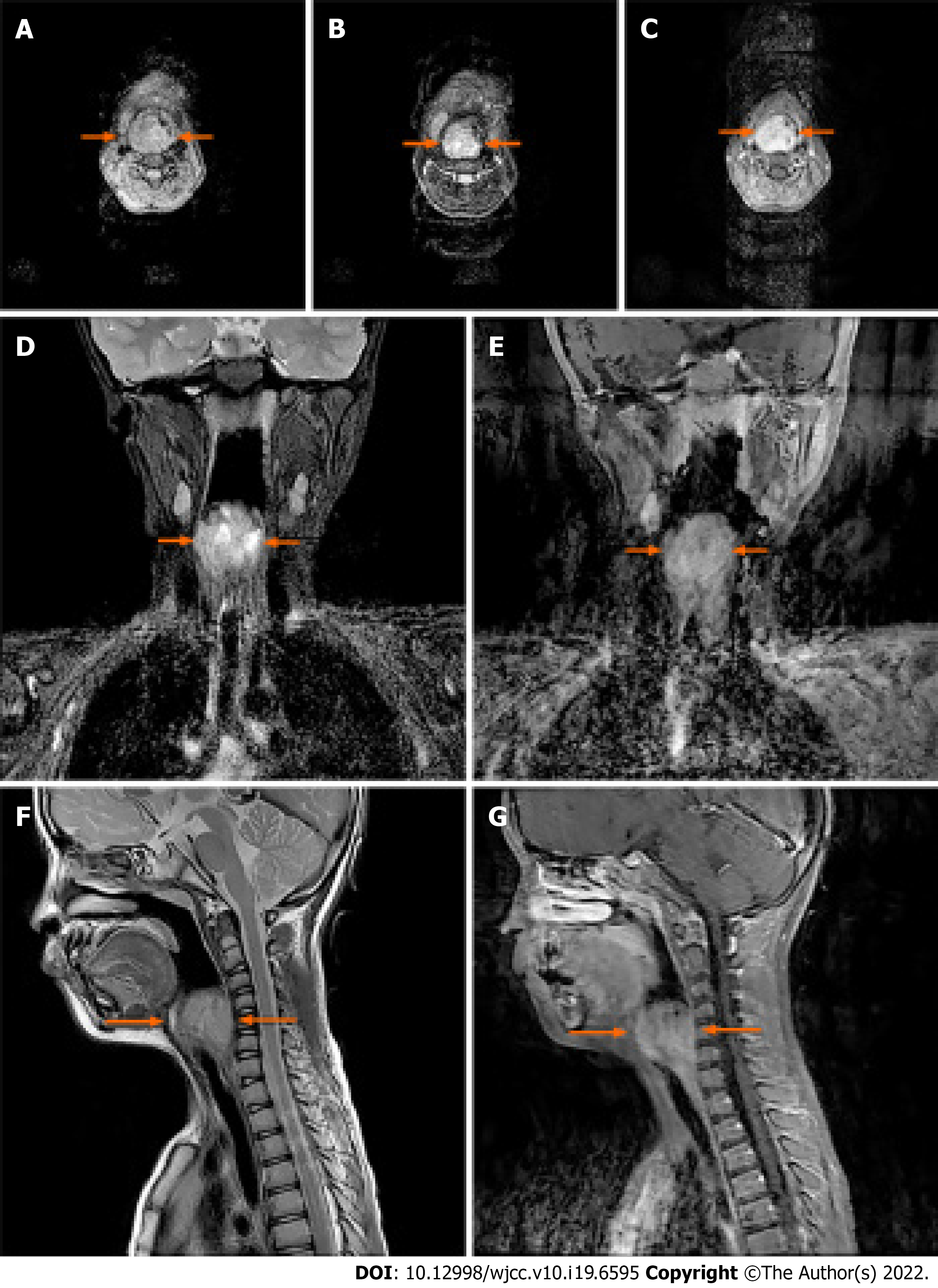
Figure 2 The soft tissue mass was indicated on the transverse plane, coronal plane and sagittal plane from 3.
0 T magnetic resonance imaging scan. A: A well-defined lesion located in the right laryngeal and piriform recess present as heterogeneous equisignal intensity on T1 -weighted image; B, D and F: High signal intensity on T2 -weighted images; C, E and G: Moreover, the contrast enhanced MRI scan displayed the prominent and heterogeneous contrast enhancement with fast perfusion mode in the early arterial phase on T1+C images.
- Citation: Chen ZH, Guo HQ, Chen JJ, Zhang Y, Zhao L. Imaging-based diagnosis for extraskeletal Ewing sarcoma in pediatrics: A case report . World J Clin Cases 2022; 10(19): 6595-6601
- URL: https://www.wjgnet.com/2307-8960/full/v10/i19/6595.htm
- DOI: https://dx.doi.org/10.12998/wjcc.v10.i19.6595


