Copyright
©The Author(s) 2022.
World J Clin Cases. Jul 6, 2022; 10(19): 6571-6579
Published online Jul 6, 2022. doi: 10.12998/wjcc.v10.i19.6571
Published online Jul 6, 2022. doi: 10.12998/wjcc.v10.i19.6571
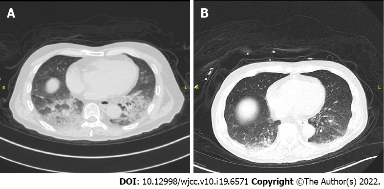
Figure 2 Chest computed tomography.
A: Diffuse exudation in the lungs; B: Exudation in the lungs was significantly cleared up. Imaging A and B were performed at an interval of approximate 3 h for excluding pulmonary embolism and aortic dissection.
- Citation: Li J, Feng ZJ, Liu L, Ma YJ. Acute methanol poisoning with bilateral diffuse cerebral hemorrhage: A case report. World J Clin Cases 2022; 10(19): 6571-6579
- URL: https://www.wjgnet.com/2307-8960/full/v10/i19/6571.htm
- DOI: https://dx.doi.org/10.12998/wjcc.v10.i19.6571


