Copyright
©The Author(s) 2022.
World J Clin Cases. Jul 6, 2022; 10(19): 6406-6416
Published online Jul 6, 2022. doi: 10.12998/wjcc.v10.i19.6406
Published online Jul 6, 2022. doi: 10.12998/wjcc.v10.i19.6406
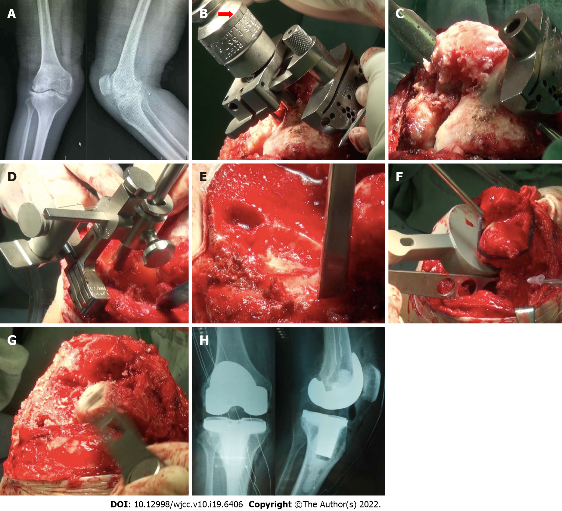
Figure 1 Process of total knee arthroplasty.
A: Preoperative X-ray of the knee joint in anterior and lateral position; B: The distal femoral valgus osteotomy angle was 7° (red arrow); C: The thickness of the medial femoral condyle osteotomy was more than that of the lateral condyle; D: Tibial plateau osteotomy was taken as the medial tibial plateau reference; E: The osteophyte of the lateral tibial plateau was removed, and the lateral plateau bone defect (black circle) was visible; F: Release of the lateral collateral ligament; G: The thickness of distal femoral osteotomy was increased because the extension space was smaller than the flexion space; H: Postoperative X-ray of the knee joint in anterior and lateral position.
- Citation: Lv SJ, Wang XJ, Huang JF, Mao Q, He BJ, Tong PJ. Total knee arthroplasty in Ranawat II valgus deformity with enlarged femoral valgus cut angle: A new technique to achieve balanced gap. World J Clin Cases 2022; 10(19): 6406-6416
- URL: https://www.wjgnet.com/2307-8960/full/v10/i19/6406.htm
- DOI: https://dx.doi.org/10.12998/wjcc.v10.i19.6406


