Copyright
©The Author(s) 2022.
World J Clin Cases. Jun 26, 2022; 10(18): 6261-6268
Published online Jun 26, 2022. doi: 10.12998/wjcc.v10.i18.6261
Published online Jun 26, 2022. doi: 10.12998/wjcc.v10.i18.6261
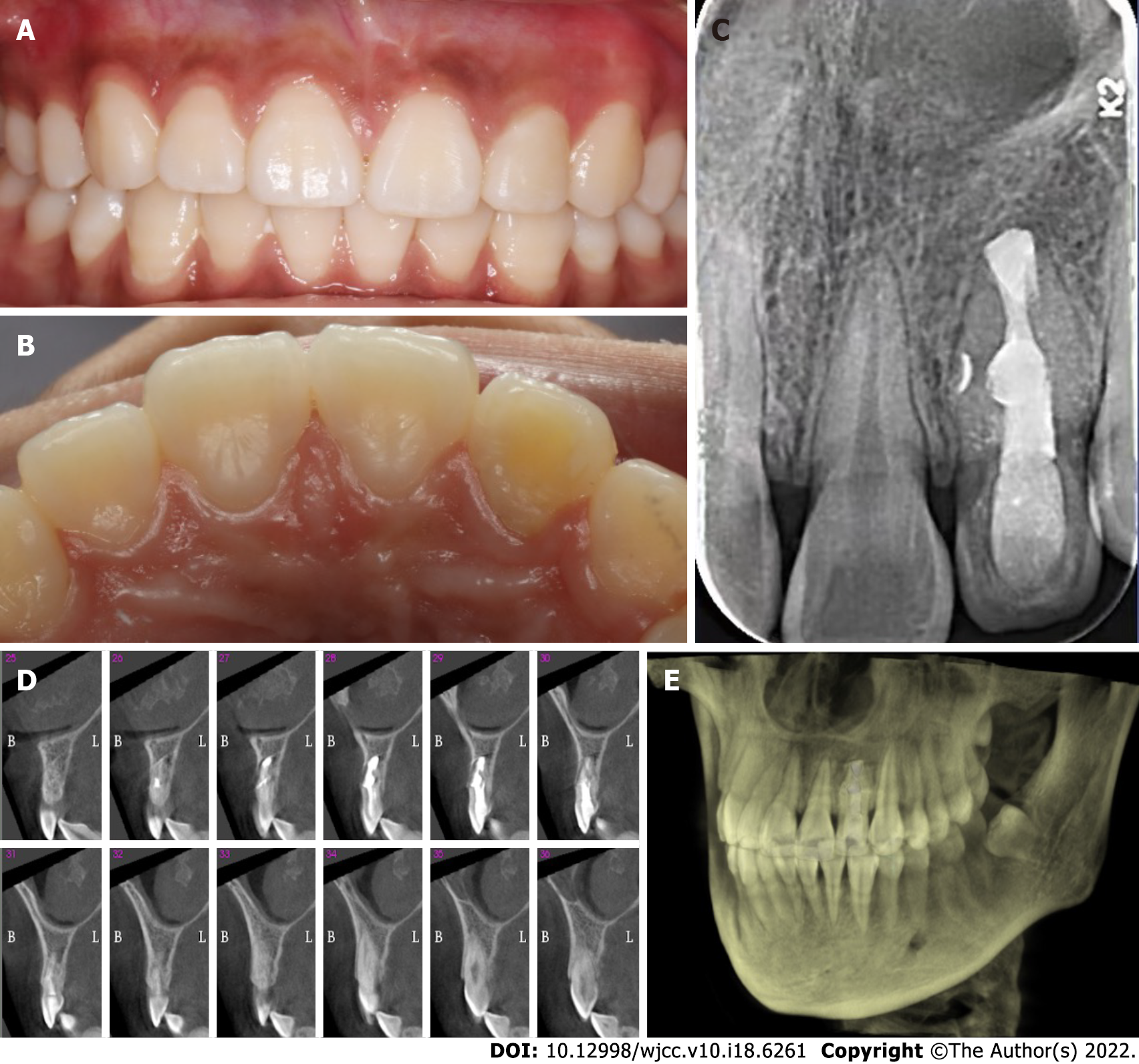
Figure 4 Follow-up photographs and radiograph.
A: Three-year recall labial view; B: Three-year recall palatal view; C: Three-year recall radiograph. The obturated lateral canal was visualized; D: Sagittal sections showing bony healing; E: Three-dimensional reconstruction indicating continuous bony plates.
- Citation: Zhang J, Li N, Li WL, Zheng XY, Li S. Management of type IIIb dens invaginatus using a combination of root canal treatment, intentional replantation, and surgical therapy: A case report. World J Clin Cases 2022; 10(18): 6261-6268
- URL: https://www.wjgnet.com/2307-8960/full/v10/i18/6261.htm
- DOI: https://dx.doi.org/10.12998/wjcc.v10.i18.6261


