Copyright
©The Author(s) 2022.
World J Clin Cases. Jun 16, 2022; 10(17): 5899-5909
Published online Jun 16, 2022. doi: 10.12998/wjcc.v10.i17.5899
Published online Jun 16, 2022. doi: 10.12998/wjcc.v10.i17.5899
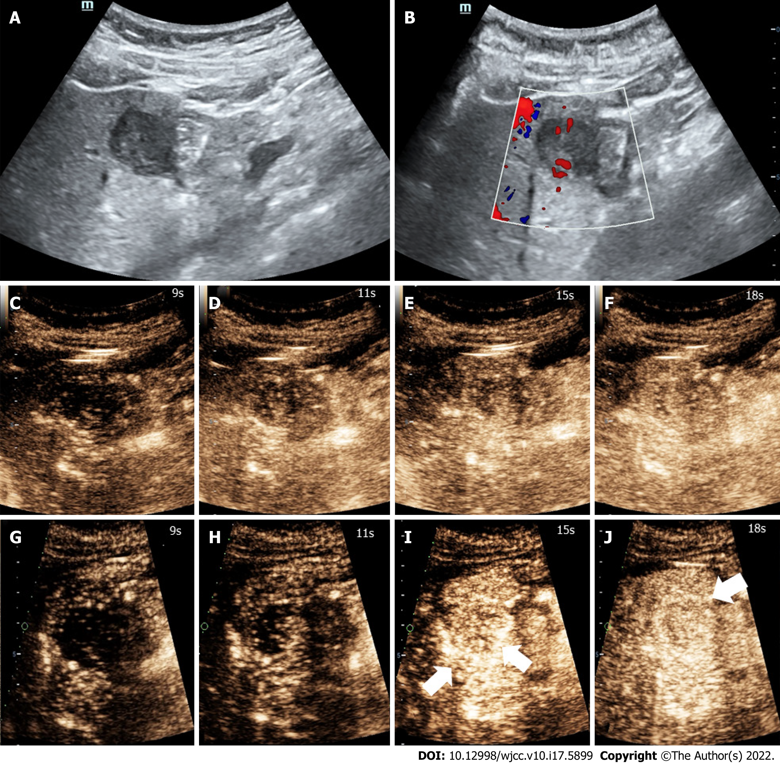
Figure 2 High-frame-rate contrast-enhanced ultrasound findings.
Images showing high-frame-rate contrast-enhanced ultrasound perfusion patterns corresponding to those in contrast-enhanced ultrasound images. Concentric enhancement was more obvious. Irregular vessel column and rim enhancement can be seen (white arrows). A: Conventional ultrasound showing an uneven hypo-echo lesion with an approximate size of 3.5 cm × 2.2 cm × 2.4 cm and a peripheral hypoechoic halo located in the left lobe of the liver; B: Color Doppler flow imaging showing the dot-linear blood flow signal within the lesion; C-E: Contrast-enhanced ultrasound findings; F: The lesion showed heterogeneous hyper-enhancement at peak; G-J: We could not find obvious concentric and rim enhancement on contrast-enhanced ultrasound.
- Citation: Chen JH, Huang Y. High-frame-rate contrast-enhanced ultrasound findings of liver metastasis of duodenal gastrointestinal stromal tumor: A case report and literature review. World J Clin Cases 2022; 10(17): 5899-5909
- URL: https://www.wjgnet.com/2307-8960/full/v10/i17/5899.htm
- DOI: https://dx.doi.org/10.12998/wjcc.v10.i17.5899


