Copyright
©The Author(s) 2022.
World J Clin Cases. Jun 16, 2022; 10(17): 5899-5909
Published online Jun 16, 2022. doi: 10.12998/wjcc.v10.i17.5899
Published online Jun 16, 2022. doi: 10.12998/wjcc.v10.i17.5899
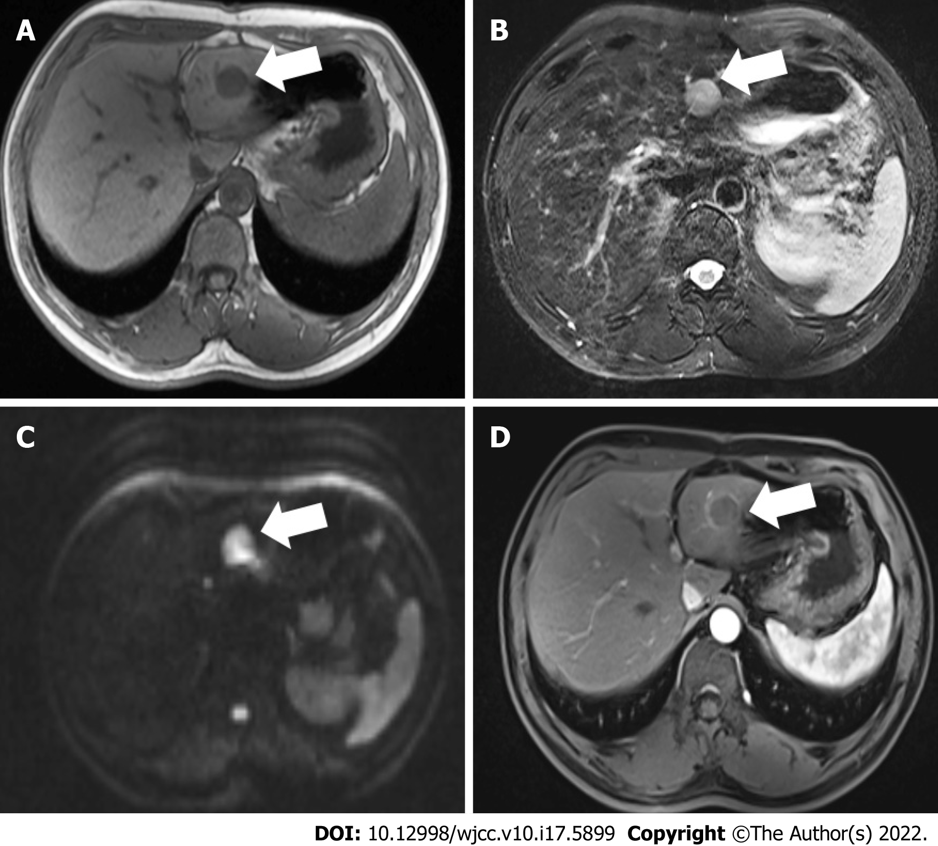
Figure 1 Magnetic resonance imaging findings.
The lesion is approximately 2.3 cm in the left external lobe. A: T1-weighted image of the nodule showing hypo-intensity; B T2-weighted image of the tumor showing higher intensity; C: Diffusion-weighted image of the tumor showing a higher intensity; D: The lesion presented edge enhancement after reinforcement (white arrows).
- Citation: Chen JH, Huang Y. High-frame-rate contrast-enhanced ultrasound findings of liver metastasis of duodenal gastrointestinal stromal tumor: A case report and literature review. World J Clin Cases 2022; 10(17): 5899-5909
- URL: https://www.wjgnet.com/2307-8960/full/v10/i17/5899.htm
- DOI: https://dx.doi.org/10.12998/wjcc.v10.i17.5899


