Copyright
©The Author(s) 2022.
World J Clin Cases. Jun 16, 2022; 10(17): 5861-5868
Published online Jun 16, 2022. doi: 10.12998/wjcc.v10.i17.5861
Published online Jun 16, 2022. doi: 10.12998/wjcc.v10.i17.5861
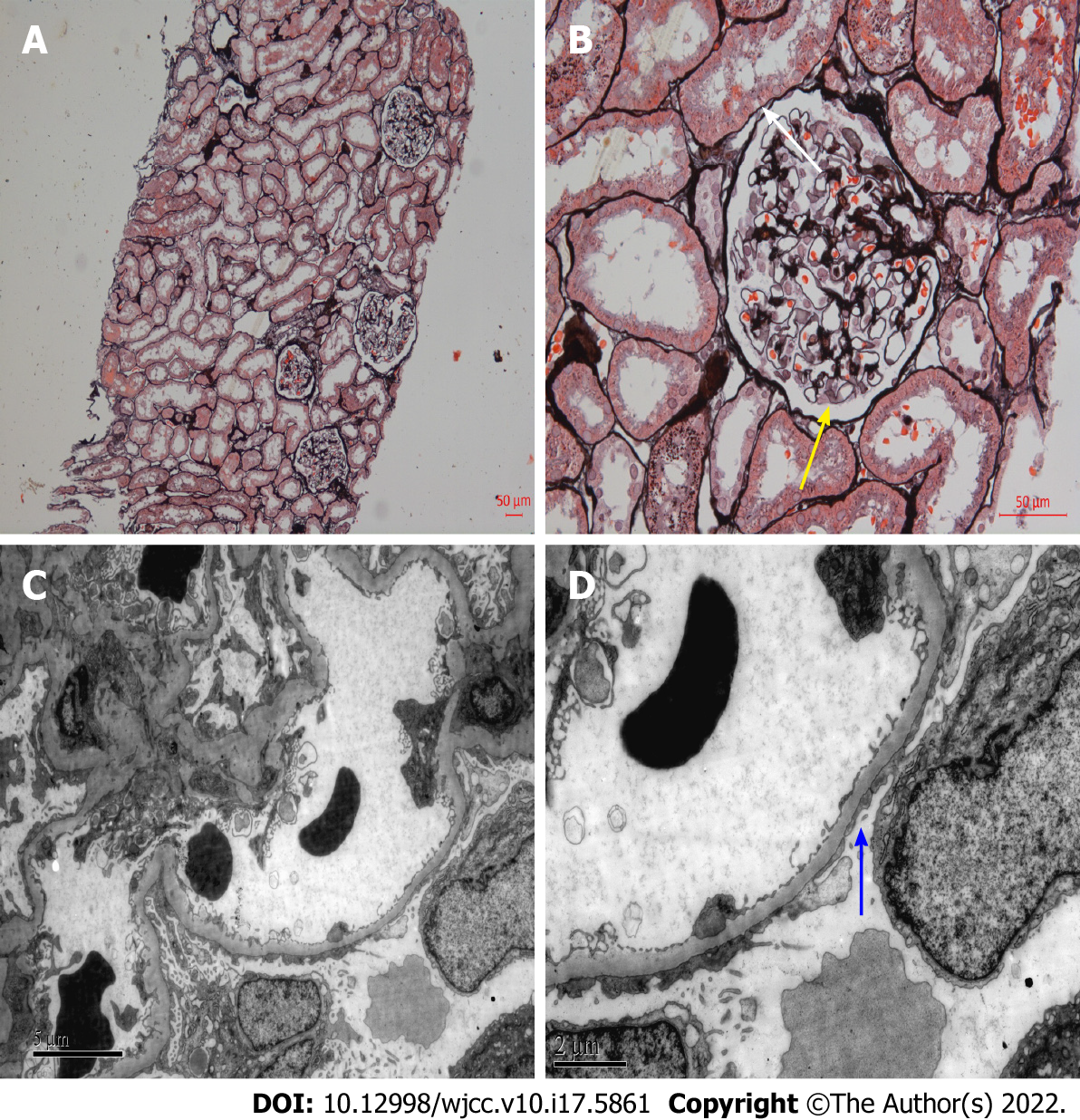
Figure 1 Light microscopy and electron microscopy of histological changes of renal biopsy 20 years ago.
A: Periodic acid-silver methenamine (PASM), × 100. No obvious lesions in renal interstitium and arterioles; B: PASM, × 400. Vacuolar degeneration of glomerular capillary basement membrane (yellow arrow), renal tubular epithelial cells vacuoles and granular degeneration (white arrow); C and D: Extensive fusion of foot process of glomerular visceral epithelial cells (C: × 6000; D: × 12000, blue arrow).
- Citation: Tang L, Cai Z, Wang SX, Zhao WJ. Transition from minimal change disease to focal segmental glomerulosclerosis related to occupational exposure: A case report. World J Clin Cases 2022; 10(17): 5861-5868
- URL: https://www.wjgnet.com/2307-8960/full/v10/i17/5861.htm
- DOI: https://dx.doi.org/10.12998/wjcc.v10.i17.5861


