Copyright
©The Author(s) 2022.
World J Clin Cases. Jun 16, 2022; 10(17): 5805-5809
Published online Jun 16, 2022. doi: 10.12998/wjcc.v10.i17.5805
Published online Jun 16, 2022. doi: 10.12998/wjcc.v10.i17.5805
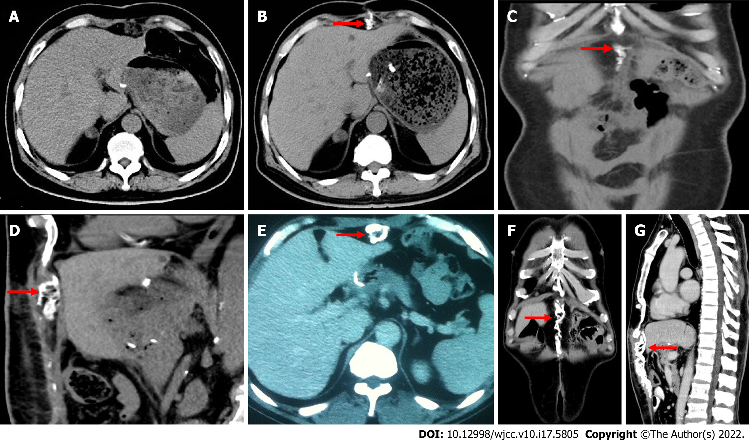
Figure 1 Imaging examinations.
A: Computed tomography (CT) scan 2 wk postoperatively in case 1 shows no obvious abnormality beneath the incision; B: CT scan 6 wk postoperatively in case 1 shows calcified tissue beneath the incision (arrow); C and D: Coronal and sagittal images of calcified tissue in case 1 between the incision and liver (arrow); E: CT scan 4 mo postoperatively in case 2 shows calcified tissue beneath the midline incision (arrow); F and G: Coronal and sagittal images in case 2 show extension of calcification (arrow).
- Citation: Zhang X, Xia PT, Ma YC, Dai Y, Wang YL. Heterotopic ossification beneath the upper abdominal incision after radical gastrectomy: Two case reports. World J Clin Cases 2022; 10(17): 5805-5809
- URL: https://www.wjgnet.com/2307-8960/full/v10/i17/5805.htm
- DOI: https://dx.doi.org/10.12998/wjcc.v10.i17.5805


