Copyright
©The Author(s) 2022.
World J Clin Cases. Jun 16, 2022; 10(17): 5732-5740
Published online Jun 16, 2022. doi: 10.12998/wjcc.v10.i17.5732
Published online Jun 16, 2022. doi: 10.12998/wjcc.v10.i17.5732
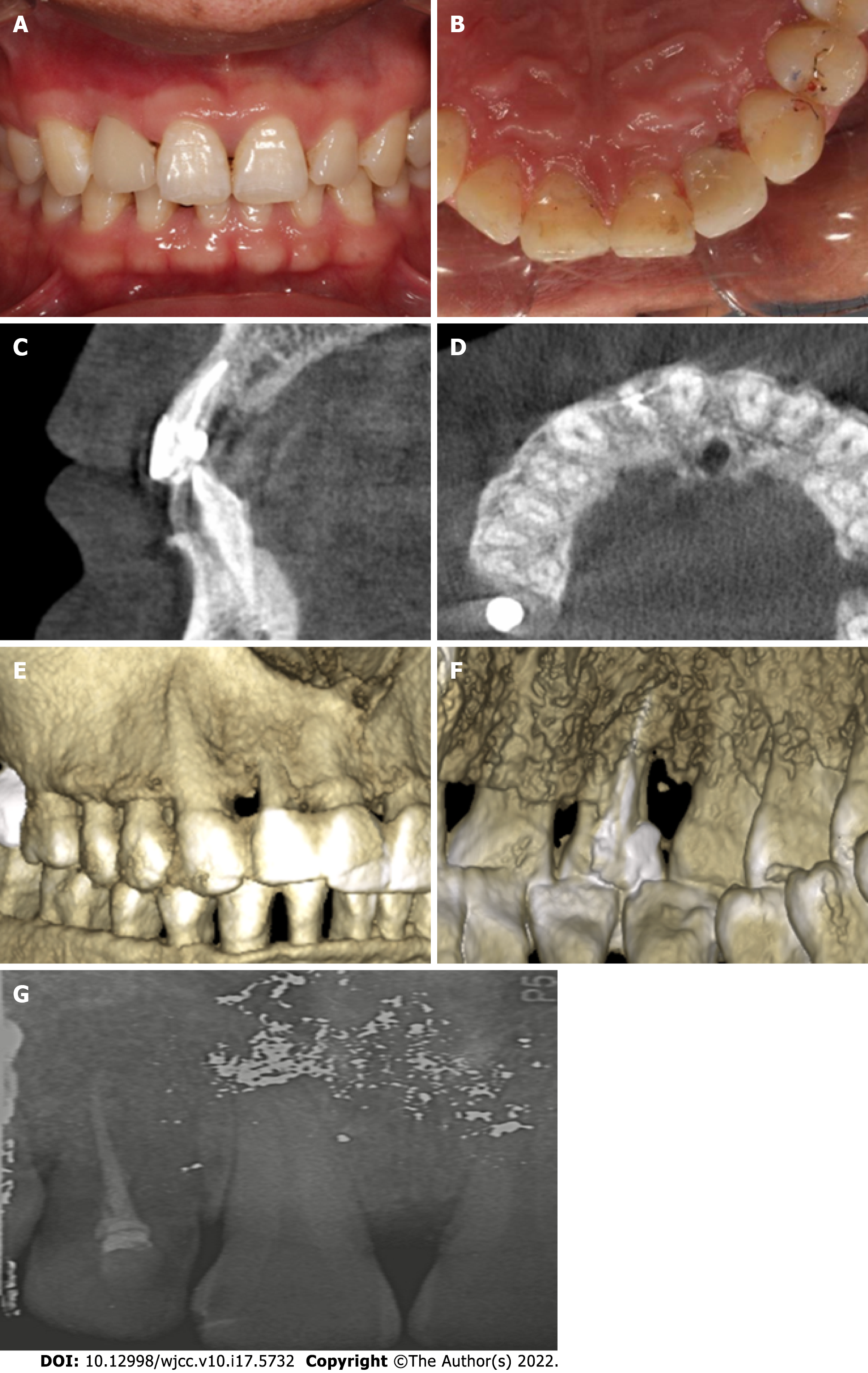
Figure 3 Two-year follow-up after surgery.
A: Buccal view of the clinical photograph after veneer preparation; B: Palatal view of the clinical photograph after veneer preparation; C: Postoperative cone beam computed tomography at 1 year showed the disappearance of diffuse radiolucency; D: Axial view of the middle third section of tooth 12 showed the filling of the bone defect around the distal aspect of the root; E: Dimensional reconstruction showed disappearance of the bone defect around tooth 12; F: Dimensional reconstruction showed that the groove was sealed; G: Periapical radiograph at the 2-year recall.
- Citation: Ling DH, Shi WP, Wang YH, Lai DP, Zhang YZ. Management of the palato-radicular groove with a periodontal regenerative procedure and prosthodontic treatment: A case report. World J Clin Cases 2022; 10(17): 5732-5740
- URL: https://www.wjgnet.com/2307-8960/full/v10/i17/5732.htm
- DOI: https://dx.doi.org/10.12998/wjcc.v10.i17.5732


