Copyright
©The Author(s) 2022.
World J Clin Cases. Jun 16, 2022; 10(17): 5708-5716
Published online Jun 16, 2022. doi: 10.12998/wjcc.v10.i17.5708
Published online Jun 16, 2022. doi: 10.12998/wjcc.v10.i17.5708
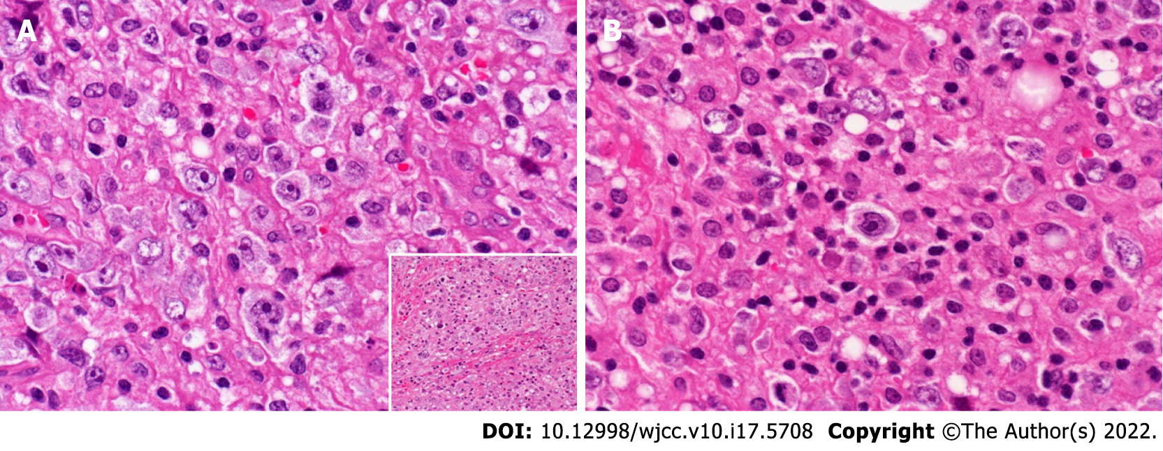
Figure 3 Large lymphoma cells in the biopsied right supraclavicular lymph node.
A: Sheet-like growth of atypical and pleomorphic cells and centroblastic cells (× 400). Inset: Fibrosis around the sheets of lymphoma cells (× 20); B: Large, atypical cells with some retracted pale cytoplasm scattered among the inflammatory cells (× 400).
- Citation: Hojo N, Nagasaki M, Mihara Y. Gray zone lymphoma effectively treated with cyclophosphamide, doxorubicin, vincristine, prednisolone, and rituximab chemotherapy: A case report. World J Clin Cases 2022; 10(17): 5708-5716
- URL: https://www.wjgnet.com/2307-8960/full/v10/i17/5708.htm
- DOI: https://dx.doi.org/10.12998/wjcc.v10.i17.5708


