Copyright
©The Author(s) 2022.
World J Clin Cases. Jun 16, 2022; 10(17): 5708-5716
Published online Jun 16, 2022. doi: 10.12998/wjcc.v10.i17.5708
Published online Jun 16, 2022. doi: 10.12998/wjcc.v10.i17.5708
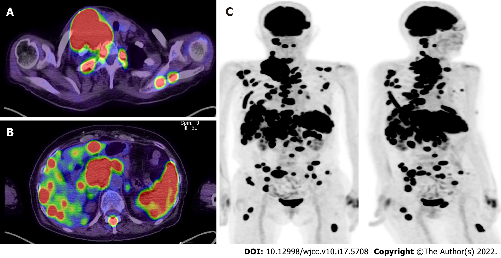
Figure 2 Fluorodeoxyglucose positron emission tomography/computed tomography revealed multiple lesions throughout the patient’s body.
A: Abnormal lymph node enlargement in the right neck, right clavicular region, and anterior mediastinum; B: Abnormal lymph node enlargement around the hepatic portal region, pancreatic head, and paraaortic region. Multiple nodular lesions in the liver and spleen; C: Multiple bone lesions in the left skull, thoracic and lumbar vertebrae, bilateral ribs, right clavicle, bilateral scapulae, lower end of the sternum, sacrum, bilateral ilia, left pubis, and bilateral femora.
- Citation: Hojo N, Nagasaki M, Mihara Y. Gray zone lymphoma effectively treated with cyclophosphamide, doxorubicin, vincristine, prednisolone, and rituximab chemotherapy: A case report. World J Clin Cases 2022; 10(17): 5708-5716
- URL: https://www.wjgnet.com/2307-8960/full/v10/i17/5708.htm
- DOI: https://dx.doi.org/10.12998/wjcc.v10.i17.5708


